12 Respiratory System

12.1 Introduction

In this chapter, you will learn about the respiratory system — the system that exchanges gases (such as oxygen and carbon dioxide) between the body and the outside air. Specifically, you will learn about:

  • The process of respiration, in which oxygen moves from the outside air into the body and carbon dioxide and other waste gases move from inside the body into the outside air.
  • The organs of the respiratory system, including the lungs, bronchial tubes, and the rest of the respiratory tract.
  • How the respiratory tract protects itself from pathogens and other potentially harmful substances in the air.
  • How the rate of breathing is regulated to maintain homeostasis of blood gases and pH.
  • How ventilation, or breathing, allows us to inhale air into the body and exhale air out of the body.
  • The conscious and unconscious control of breathing.
  • Nasal breathing compared to mouth breathing.
  • How gas exchange occurs between the air and blood in the alveoli of the lungs, and between the blood and cells throughout the body.
  • Disorders of the respiratory system, including asthma, pneumonia, chronic obstructive pulmonary disease (COPD), and lung cancer.
  • The negative health effects of smoking.

12.2 STRUCTURE AND FUNCTION OF THE RESPIRATORY SYSTEM


13.2.1 Exhale
Figure 12.1 Every breath you take… 

SEEING YOUR BREATH

Why can you “see your breath” on a cold day? The air you exhale through your nose and mouth is warm like the inside of your body. Exhaled air also contains a lot of water vapor, because it passes over moist surfaces from the lungs to the nose or mouth. The water vapor in your breath cools suddenly when it reaches the much colder outside air. This causes the water vapor to condense into a fog of tiny droplets of liquid water. You release water vapor and other gases from your body through the process of respiration.

WHAT IS RESPIRATION?

Respiration is the life-sustaining process in which gases are exchanged between the body and the outside atmosphere. Specifically, oxygen moves from the outside air into the body; and water vapor, carbon dioxide, and other waste gases move from inside the body to the outside air. Respiration is carried out mainly by the respiratory system. It is important to note that respiration by the respiratory system is not the same process as cellular respiration —which occurs inside cells — although the two processes are closely connected. Cellular respiration is the metabolic process in which cells obtain energy, usually by “burning” glucose in the presence of oxygen. When cellular respiration is aerobic, it uses oxygen and releases carbon dioxide as a waste product. Respiration by the respiratory system supplies the oxygen needed by cells for aerobic cellular respiration and removes the carbon dioxide produced by cells during cellular respiration.

Respiration by the respiratory system actually involves two subsidiary processes. One process is ventilation, or breathing. Ventilation is the physical process of conducting air to and from the lungs. The other process is gas exchange. This is the biochemical process in which oxygen diffuses out of the air and into the blood, while carbon dioxide and other waste gases diffuse out of the blood and into the air. All of the organs of the respiratory system are involved in breathing, but only the lungs are involved in gas exchange.

RESPIRATORY ORGANS

The organs of the respiratory system form a continuous system of passages, called the respiratory tract, through which air flows into and out of the body. The respiratory tract has two major divisions: the upper respiratory tract and the lower respiratory tract. The organs in each division are shown in Figure 12.2. In addition to these organs, certain muscles of the thorax (body cavity that fills the chest) are also involved in respiration by enabling breathing. Most important is a large muscle called the diaphragm, which lies below the lungs and separates the thorax from the abdomen. Smaller muscles between the ribs also play a role in breathing.

13.2.2 Respiratory Tract
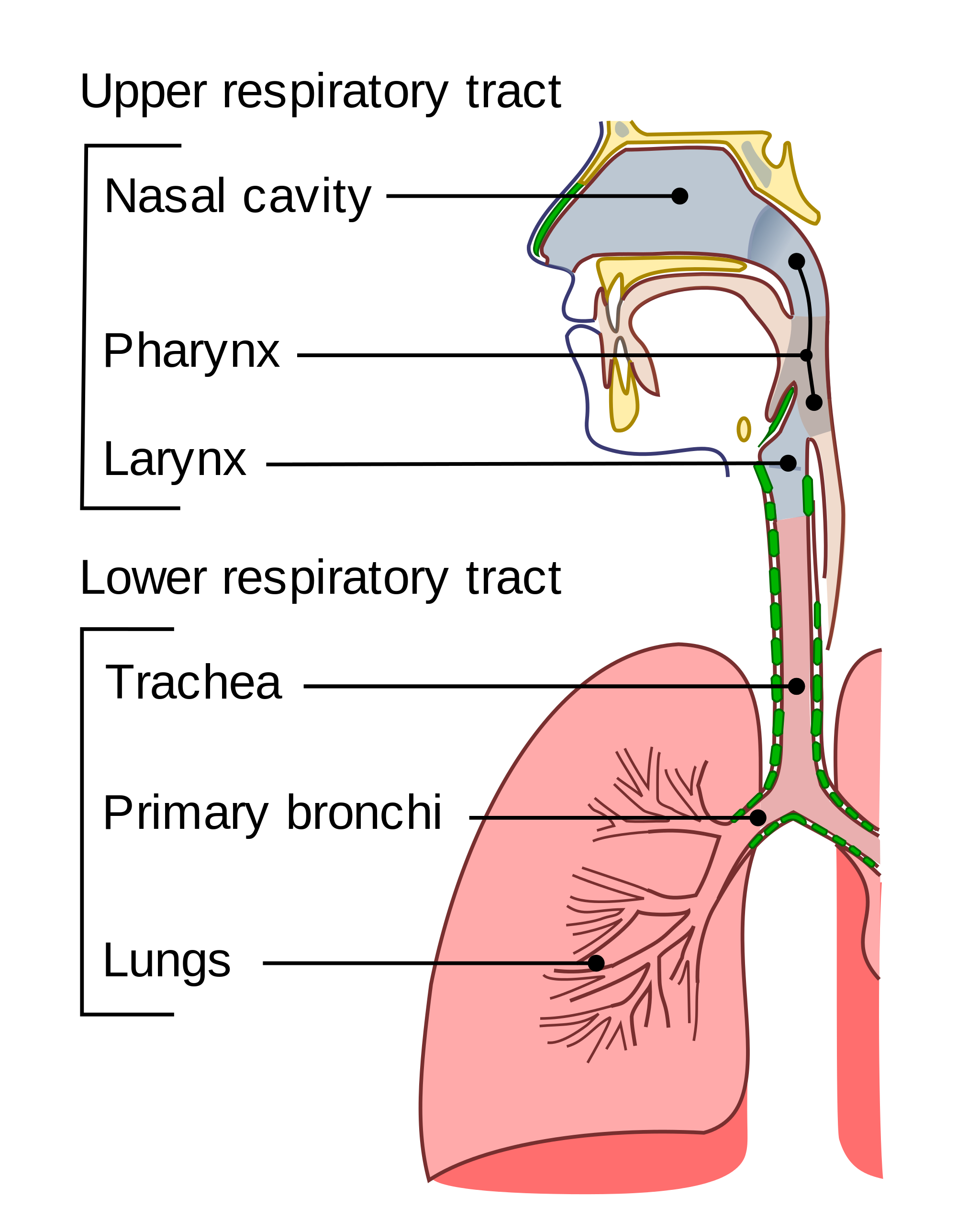
Figure 12.2 During breathing, inhaled air enters the body through the nose and passes through the respiratory tract to the lungs. Exhaled air travels from the lungs in the opposite direction.

UPPER RESPIRATORY TRACT

All of the organs and other structures of the upper respiratory tract are involved in conduction, or the movement of air into and out of the body. Upper respiratory tract organs provide a route for air to move between the outside atmosphere and the lungs. They also clean, humidify, and warm the incoming air. No gas exchange occurs in these organs.

Nasal Cavity

The nasal cavity is a large, air-filled space in the skull above and behind the nose in the middle of the face. It is a continuation of the two nostrils. As inhaled air flows through the nasal cavity, it is warmed and humidified by blood vessels very close to the surface of this epithelial tissue. Hairs in the nose and mucous produced by mucous membranes help trap larger foreign particles in the air before they go deeper into the respiratory tract. In addition to its respiratory functions, the nasal cavity also contains chemoreceptors needed for sense of smell, and contribution to the sense of taste.

Pharynx

The pharynx is a tube-like structure that connects the nasal cavity and the back of the mouth to other structures lower in the throat, including the larynx. The pharynx has dual functions — both air and food (or other swallowed substances) pass through it, so it is part of both the respiratory and the digestive systems. Air passes from the nasal cavity through the pharynx to the larynx (as well as in the opposite direction). Food passes from the mouth through the pharynx to the esophagus.

Larynx

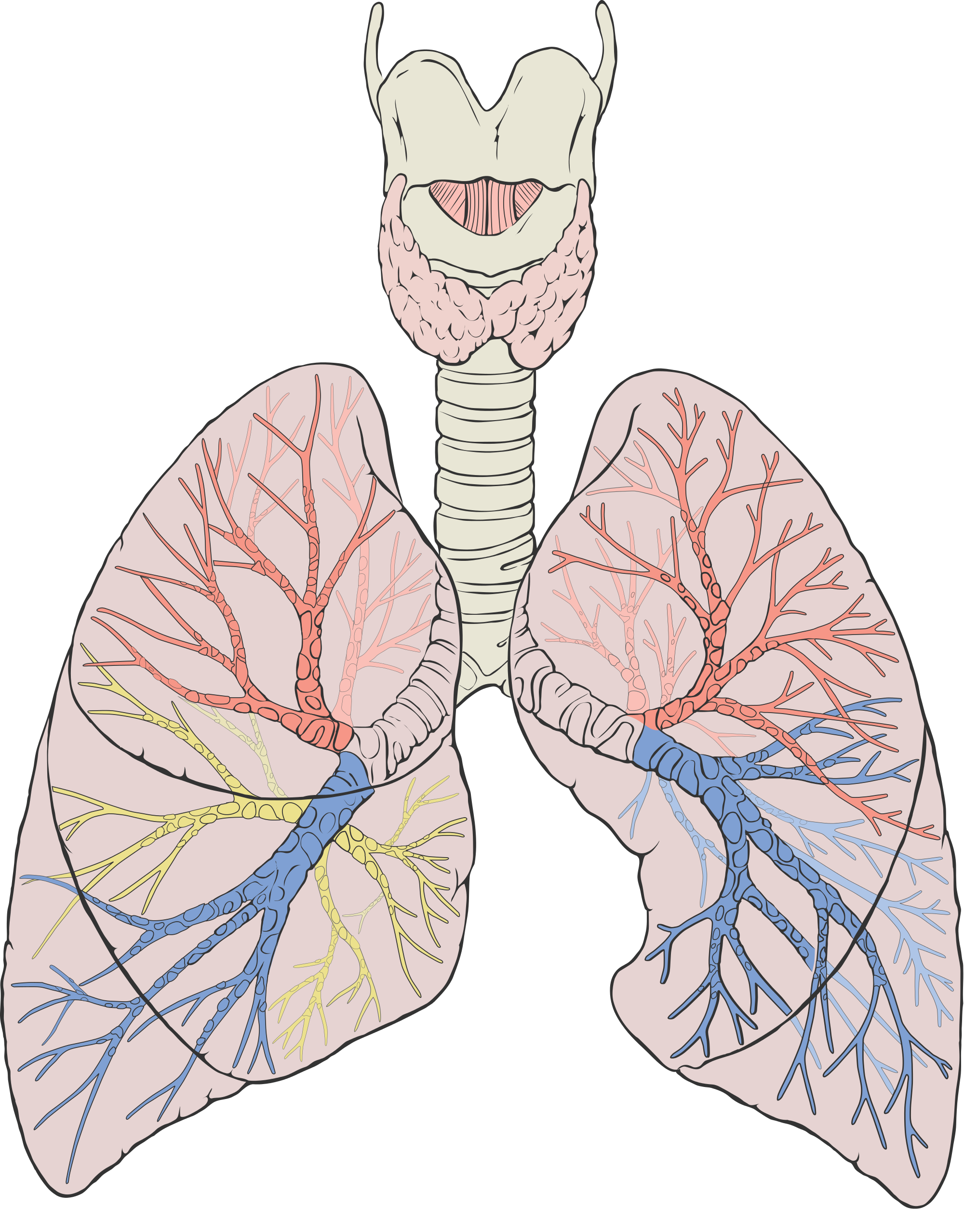
The larynx connects the pharynx and trachea, and helps to conduct air through the respiratory tract. The larynx is also called the voice box, because it contains the vocal cords, which vibrate when air flows over them, thereby producing sound. You can see the vocal cords in the larynx in Figures 12.3 and 12.4. Certain muscles in the larynx move the vocal cords apart to allow breathing. Other muscles in the larynx move the vocal cords together to allow the production of vocal sounds. The latter muscles also control the pitch of sounds and help control their volume.

A very important function of the larynx is protecting the trachea from aspirated food. When swallowing occurs, the backward motion of the tongue forces a flap called the epiglottis to close over the entrance to the larynx. (You can see the epiglottis in both Figure 12.3 and 12.4.) This prevents swallowed material from entering the larynx and moving deeper into the respiratory tract. If swallowed material does start to enter the larynx, it irritates the larynx and stimulates a strong cough reflex. This generally expels the material out of the larynx, and into the throat.

13.2.3 Larynx external view                    13.2.4 Larynx top view

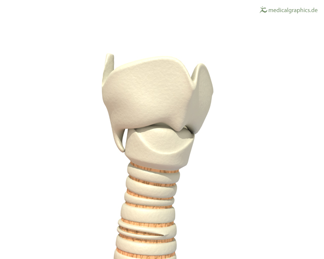
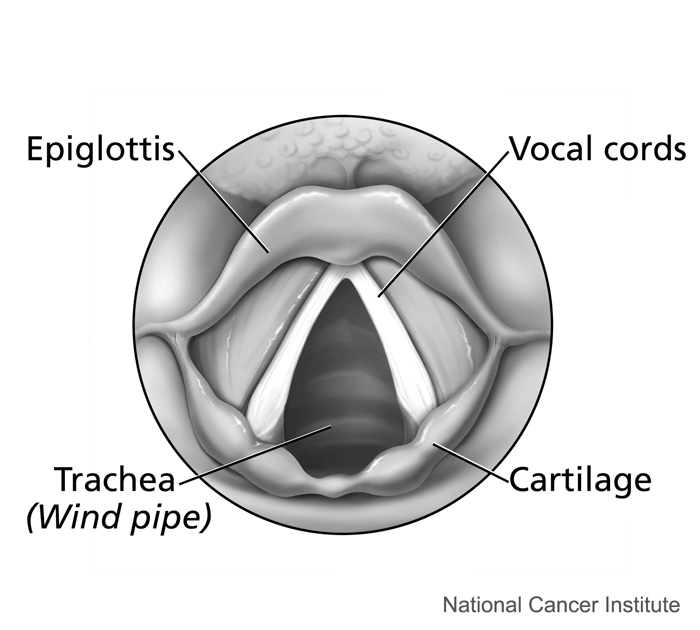
Figure 12.3 The larynx as viewed from externally.      Figure 12.4 The larynx as viewed from the top.

Larynx Model – Respiratory System, Dr. Lotz, 2018.

 LOWER RESPIRATORY TRACT


13.2.5 Branching in the lower respiratory tract
Figure 12.5 This diagram illustrates the tree-like branching of the passages of the lower respiratory tract within the lungs.

The trachea and other passages of the lower respiratory tract conduct air between the upper respiratory tract and the lungs. These passages form an inverted tree-like shape (Figure 12.5), with repeated branching as they move deeper into the lungs. All told, there are an astonishing 2,414 kilometers (1,500 miles) of airways conducting air through the human respiratory tract! It is only in the lungs, however, that gas exchange occurs between the air and the bloodstream.

Trachea

The trachea, or windpipe, is the widest passageway in the respiratory tract. It is about 2.5 cm wide and 10-15 cm long (approximately 1 inch wide and 4–6 inches long). It is formed by rings of cartilage, which make it relatively strong and resilient. The trachea connects the larynx to the lungs for the passage of air through the respiratory tract. The trachea branches at the bottom to form two bronchial tubes.

Bronchi and Bronchioles

There are two main bronchial tubes, or bronchi (singular, bronchus), called the right and left bronchi. The bronchi carry air between the trachea and lungs. Each bronchus branches into smaller, secondary bronchi; and secondary bronchi branch into still smaller tertiary bronchi. The smallest bronchi branch into very small tubules called bronchioles. The tiniest bronchioles end in alveolar ducts, which terminate in clusters of minuscule air sacs, called alveoli(singular, alveolus), in the lungs.

Lungs

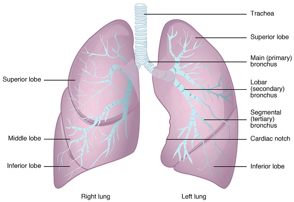
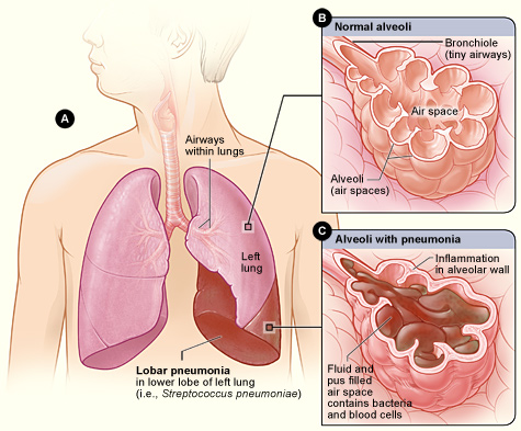
The lungs are the largest organs of the respiratory tract. They are suspended within the pleural cavity of the thorax. The lungs are surrounded by two thin membranes called pleura, which secrete fluid that allows the lungs to move freely within the pleural cavity. This is necessary so the lungs can expand and contract during breathing. In Figure 13.2.6, you can see that each of the two lungs is divided into sections. These are called lobes, and they are separated from each other by connective tissues. The right lung is larger and contains three lobes. The left lung is smaller and contains only two lobes. The smaller left lung allows room for the heart, which is just left of the center of the chest.

13.2.6 Anatomy of the Lung
Figure 12.6 The lungs are separated into the right and left lung.

As mentioned previously, the bronchi terminate in bronchioles which feed air into alveoli, tiny sacs of simple squamous epithelial tissue which make up the bulk of the lung.  The cross-section of lung tissue in the diagram below (Figure 12.7) shows the alveoli, in which gas exchange takes place with the capillary network that surrounds them.


13.2.7 Alveoli Structure
Figure 12.7 Alveoli make up the bulk of the lung and form millions of grape-like clusters of air sacs for the purpose of exchanging gases with capillaries of the cardiovascular system.
13.2.8 Alveolus
Figure 12.8 An alveolus in which gas exchange takes place with the capillary network that surrounds it. Surfactant is a liquid that covers the inside of the alveoli and prevents them from collapsing and sticking together when air empties out of them during exhalation.


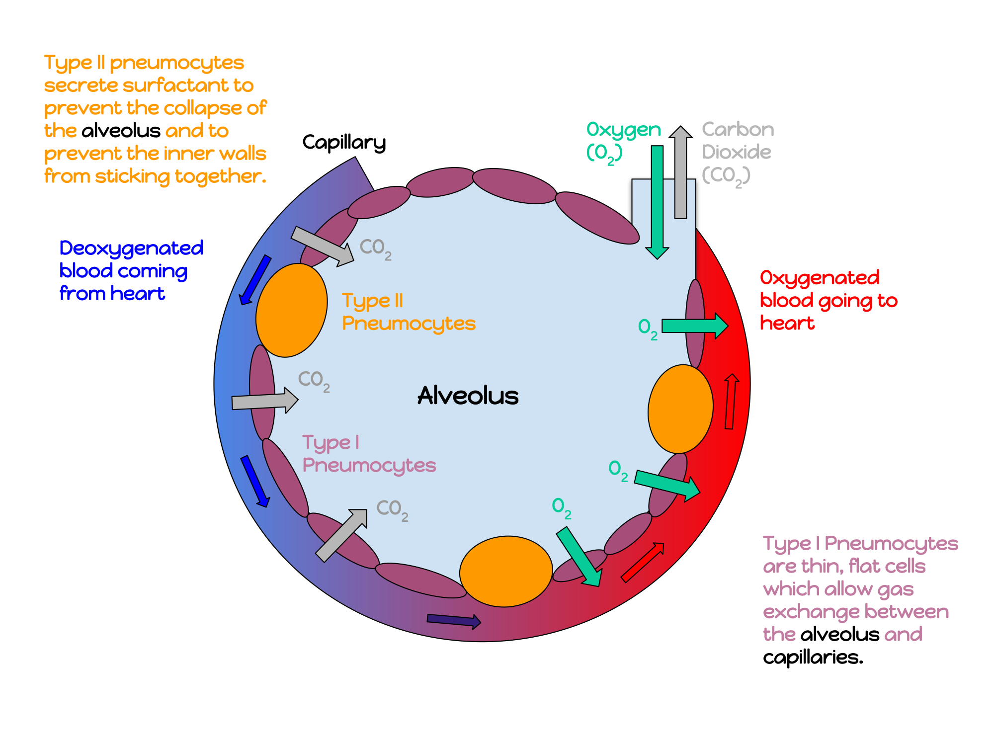
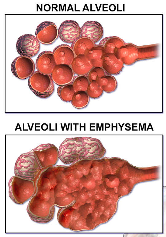
Lung tissue consists mainly of alveoli (see Figures 12.7 and 12.8). These tiny air sacs are the functional units of the lungs where gas exchange takes place. The two lungs may contain as many as 700 million alveoli, providing a huge total surface area for gas exchange to take place. In fact, alveoli in the two lungs provide as much surface area as half a tennis court! Each time you breathe in, the alveoli fill with air, making the lungs expand. Oxygen in the air inside the alveoli is absorbed by the blood via diffusion in the mesh-like network of tiny capillaries that surrounds each alveolus. The blood in these capillaries also releases carbon dioxide (also by diffusion) into the air inside the alveoli. Each time you breathe out, air leaves the alveoli and rushes into the outside atmosphere, carrying waste gases with it.

The lungs receive blood from two major sources. They receive deoxygenated blood from the right side of the heart. This blood absorbs oxygen in the lungs and carries it back to the left side heart to be pumped to cells throughout the body. The lungs also receive oxygenated blood from the heart that provides oxygen to the cells of the lungs for cellular respiration.

PROTECTING THE RESPIRATORY SYSTEM

You may be able to survive for weeks without food and for days without water, but you can survive without oxygen for only a matter of minutes — except under exceptional circumstances — so protecting the respiratory system is vital. Ensuring that a patient has an open airway is the first step in treating many medical emergencies. Fortunately, the respiratory system is well protected by the ribcage of the skeletal system. The extensive surface area of the respiratory system, however, is directly exposed to the outside world and all its potential dangers in inhaled air. It should come as no surprise that the respiratory system has a variety of ways to protect itself from harmful substances, such as dust and pathogens in the air.

The main way the respiratory system protects itself is called the mucociliary escalator. From the nose through the bronchi, the respiratory tract is covered in epithelium that contains mucus-secreting goblet cells. The mucus traps particles and pathogens in the incoming air. The epithelium of the respiratory tract is also covered with tiny cell projections called cilia (singular, cilium), as shown in the animation. The cilia constantly move in a sweeping motion upward toward the throat, moving the mucus and trapped particles and pathogens away from the lungs and toward the outside of the body. The upward sweeping motion of cilia lining the respiratory tract helps keep it free from dust, pathogens, and other harmful substances.

Mucociliary clearance, I-Hsun Wu, 2015.


13.2.9 Sneeze
Figure 12.9 Sneezing results in tiny particles from the mouth being forcefully ejected into the air.

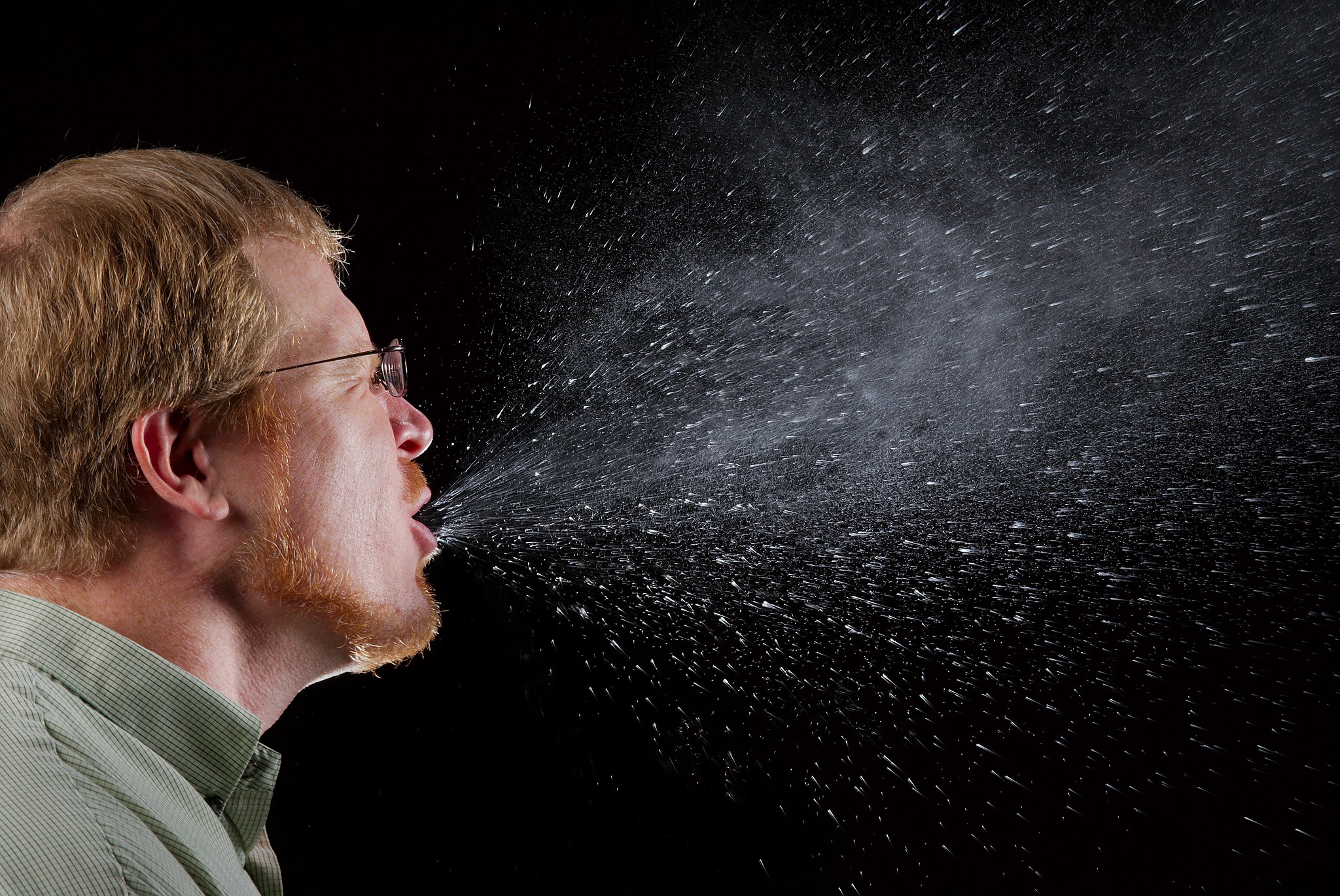
Sneezing is a similar involuntary response that occurs when nerves lining the nasal passage are irritated. It results in forceful expulsion of air from the mouth, which sprays millions of tiny droplets of mucus and other debris out of the mouth and into the air, as shown in Figure 12.9. This explains why it is so important to sneeze into a tissue (rather than the air) if we are to prevent the transmission of respiratory pathogens.


How the Respiratory System Works with Other Organ Systems

The amount of oxygen and carbon dioxide in the blood must be maintained within a limited range for survival of the organism. Cells cannot survive for long without oxygen, and if there is too much carbon dioxide in the blood, the blood becomes dangerously acidic (pH is too low). Conversely, if there is too little carbon dioxide in the blood, the blood becomes too basic (pH is too high). The respiratory system works hand-in-hand with the nervous and cardiovascular systems to maintain homeostasis in blood gases and pH.

It is the level of carbon dioxide — rather than the level of oxygen — that is most closely monitored to maintain blood gas and pH homeostasis. The level of carbon dioxide in the blood is detected by cells in the brain, which speed up or slow down the rate of breathing through the autonomic nervous system as needed to bring the carbon dioxide level within the normal range. Faster breathing lowers the carbon dioxide level (and raises the oxygen level and pH), while slower breathing has the opposite effects. In this way, the levels of carbon dioxide, oxygen, and pH are maintained within normal limits.

The respiratory system also works closely with the cardiovascular system to maintain homeostasis. The respiratory system exchanges gases with the outside air, but it needs the cardiovascular system to carry them to and from body cells. Oxygen is absorbed by the blood in the lungs and then transported through a vast network of blood vessels to cells throughout the body, where it is needed for aerobic cellular respiration. The same system absorbs carbon dioxide from cells and carries it to the respiratory system for removal from the body.

FEATURE: MY HUMAN BODY

Choking due to a foreign object becoming lodged in the airway results in nearly 5 thousand deaths in Canada each year. In addition, choking accounts for almost 40% of unintentional injuries in infants under the age of one.  For the sake of your own human body, as well as those of loved ones, you should be aware of choking risks, signs, and treatments.

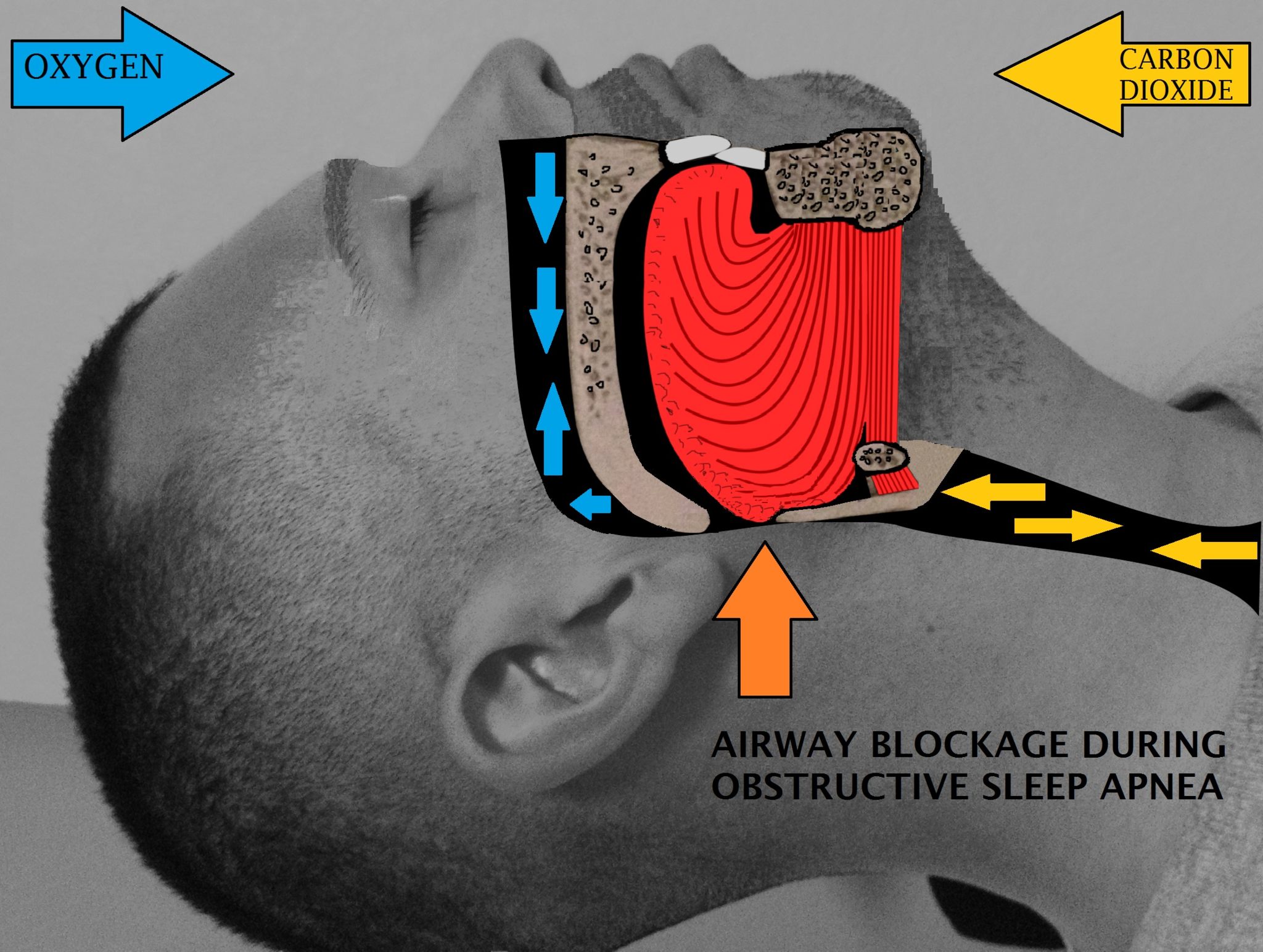
Choking is the mechanical obstruction of the flow of air from the atmosphere into the lungs. It prevents breathing, and may be partial or complete. Partial choking allows some — though inadequate — air flow into the lungs. Prolonged or complete choking results in asphyxia, or suffocation, which is potentially fatal.

Obstruction of the airway typically occurs in the pharynx or trachea. Young children are more prone to choking than are older people, in part because they often put small objects in their mouth and do not understand the risk of choking that they pose. Young children may choke on small toys or parts of toys, or on household objects, in addition to food. Foods that are round (hotdogs, carrots, grapes) or can adapt their shape to that of the pharynx (bananas, marshmallows), are especially dangerous, and may cause choking in adults, as well as children.

How can you tell if a loved one is choking? The person cannot speak or cry out, or has great difficulty doing so. Breathing, if possible, is labored, producing gasping or wheezing. The person may desperately clutch at his or her throat or mouth. If breathing is not soon restored, the person’s face will start to turn blue from lack of oxygen. This will be followed by unconsciousness, brain damage, and possibly death if oxygen deprivation continues beyond a few minutes.

If an infant is choking, turning the baby upside down and slapping him on the back may dislodge the obstructing object. To help an older person who is choking, first encourage the person to cough. Give them a few hard back slaps to help force the lodged object out of the airway. If these steps fail, perform the Heimlich maneuver on the person. See the series of videos below, from ProCPR, which demonstrate several ways to help someone who is choking based on age.


Conscious Adult Choking, ProCPR, 2016.

Conscious Child Choking, ProCPR, 2009.

Conscious Infant Choking, ProCPR, 2011

Review

  1. What is respiration, as carried out by the respiratory system? Name the two subsidiary processes it involves.
  2. Describe the respiratory tract.
  3. Identify the organs of the upper respiratory tract. What are their functions?
  4. List the organs of the lower respiratory tract. Which organs are involved only in conduction?
  5. Where does gas exchange take place?
  6. How does the respiratory system protect itself from potentially harmful substances in the air?
  7. Explain how the rate of breathing is controlled.
  8. Why does the respiratory system need the cardiovascular system to help it perform its main function of gas exchange?
  9. Describe two ways in which the body prevents food from entering the lungs.
  10. What is the relationship between respiration and cellular respiration?

How do lungs work? – Emma Bryce, TED-Ed, 2014.

12.3 BREATHING

13.3.1 Butterfly Stroke
Figure 12.10 How long can you hold your breath?

DOING THE ‘FLY

The swimmer in the Figure 12.10 photo is doing the butterfly stroke, a swimming style that requires the swimmer to carefully control his breathing, so it is coordinated with his swimming movements. Breathing is the process of moving air into and out of the lungs, which are the organs in which gas exchange takes place between the atmosphere and the body. Breathing is also called ventilation, and it is one of two parts of the life-sustaining process of respiration. The other part is gas exchange. Before you can understand how breathing is controlled, you need to know how breathing occurs.

HOW BREATHING OCCURS

Breathing is a two-step process that includes drawing air into the lungs, or inhaling, and letting air out of the lungs, or exhaling. Both processes are illustrated in Figure 12.11


13.3.2 Inhalation and Exhalation
Figure 12.11 Breathing depends mainly on repeated contractions of the diaphragm.

INHALING

Inhaling is an active process that results mainly from contraction of a muscle called the diaphragm, shown in Figure 12.11. The diaphragmis a large, dome-shaped muscle below the lungs that separates the thoracic (chest) and abdominal cavities. When the diaphragm contracts it moves down causing the thoracic cavity to expand, and the contents of the abdomen to be pushed downward. Other muscles — such as intercostal muscles between the ribs — also contribute to the process of inhalation, especially when inhalation is forced, as when taking a deep breath. These muscles help increase thoracic volume by expanding the ribs outward. The increase in thoracic volume creates a decrease in thoracic air pressure.  With the chest expanded, there is lower air pressure inside the lungs than outside the body, so outside air flows into the lungs via the respiratory tract according the the pressure gradient (high pressure flows to lower pressure).

EXHALING

Exhaling involves the opposite series of events. The diaphragm relaxes, so it moves upward and decreases the volume of the thorax. Air pressure inside the lungs increases, so it is higher than the air pressure outside the lungs. Exhalation, unlike inhalation, is typically a passive process that occurs mainly due to the elasticity of the lungs. With the change in air pressure, the lungs contract to their pre-inflated size, forcing out the air they contain in the process. Air flows out of the lungs, similar to the way air rushes out of a balloon when it is released. If exhalation is forced, internal intercostal and abdominal muscles may help move the air out of the lungs.

CONTROL OF BREATHING

Breathing is one of the few vital bodily functions that can be controlled consciously, as well as unconsciously. Think about using your breath to blow up a balloon. You take a long, deep breath, and then you exhale the air as forcibly as you can into the balloon. Both the inhalation and exhalation are consciously controlled.

CONSCIOUS CONTROL OF BREATHING

You can control your breathing by holding your breath, slowing your breathing, or hyperventilating, which is breathing more quickly and shallowly than necessary. You can also exhale or inhale more forcefully or deeply than usual. Conscious control of breathing is common in many activities besides blowing up balloons, including swimming, speech training, singing, playing many different musical instruments (Figure 12.12), and doing yoga, to name just a few.


13.3.3 Conscious Control of Breathing
Figure 12.12 Playing the trumpet is hard work. Exhaled air must be forced through the lips hard enough to create a vibrating column of air inside the instrument.

There are limits on the conscious control of breathing. For example, it is not possible for a healthy person to voluntarily stop breathing indefinitely. Before long, there is an irrepressible urge to breathe. If you were able to stop breathing for a long enough time, you would lose consciousness. The same thing would happen if you were to hyperventilate for too long. Once you lose consciousness so you can no longer exert conscious control over your breathing, involuntary control of breathing takes over.

UNCONSCIOUS CONTROL OF BREATHING

Unconscious breathing is controlled by respiratory centers in the medulla and pons of the brainstem (see Figure 13.3.4). The respiratory centers automatically and continuously regulate the rate of breathing based on the body’s needs. These are determined mainly by blood acidity, or pH. When you exercise, for example, carbon dioxide levels increase in the blood, because of increased cellular respiration by muscle cells. The carbon dioxide reacts with water in the blood to produce carbonic acid, making the blood more acidic, so pH falls. The drop in pH is detected by chemoreceptors in the medulla. Blood levels of oxygen and carbon dioxide, in addition to pH, are also detected by chemoreceptors in major arteries, which send the “data” to the respiratory centers. The latter respond by sending nerve impulses to the diaphragm, “telling” it to contract more quickly so the rate of breathing speeds up. With faster breathing, more carbon dioxide is released into the air from the blood, and blood pH returns to the normal range.

13.3.4 Nervous Control of Respiration
Figure 12.13 Clusters of cells in the pons and medulla of the brain stem are the respiratory centers of the brain that have involuntary control over breathing.

The opposite events occur when the level of carbon dioxide in the blood becomes too low, and blood pH rises. This may occur with involuntary hyperventilation, which can happen in panic attacks, episodes of severe pain, asthma attacks, and many other situations. When you hyperventilate, you blow off a lot of carbon dioxide, leading to a drop in blood levels of carbon dioxide. The blood becomes more basic (alkaline), causing its pH to rise.

NASAL VS. MOUTH BREATHING

Nasal breathing is breathing through the nose rather than the mouth, and it is generally considered to be superior to mouth breathing. The hair-lined nasal passages do a better job of filtering particles out of the air before it moves deeper into the respiratory tract. The nasal passages are also better at warming and moistening the air, so nasal breathing is especially advantageous in the winter when the air is cold and dry. In addition, the smaller diameter of the nasal passages creates greater pressure in the lungs during exhalation. This slows the emptying of the lungs, giving them more time to extract oxygen from the air.

FEATURE: MYTH VS. REALITY

Drowning is defined as respiratory impairment from being in or under a liquid. It is further classified according to its outcome into: death, ongoing health problems, or no ongoing health problems (full recovery). Four hundred Canadians die annually from drowning, and drowning is one of the leading causes of death in children under the age of five. There are some potentially dangerous myths about drowning, and knowing what they are might save your life or the life of a loved one, especially a child.

Myth Reality
“People drown when they aspirate water into their lungs.” Generally, in the early stages of drowning, very little water enters the lungs. A small amount of water entering the trachea causes a muscular spasm in the larynx that seals the airway and prevents the passage of water into the lungs. This spasm is likely to last until unconsciousness occurs.
“You can tell when someone is drowning because they will shout for help and wave their arms to attract attention.” The muscular spasm that seals the airway prevents the passage of air, as well as water, so a person who is drowning is unable to shout or call for help. In addition, instinctive reactions that occur in the final minute or so before a drowning person sinks under the water may look similar to calm, safe behavior. The head is likely to be low in the water, tilted back, with the mouth open. The person may have uncontrolled movements of the arms and legs, but they are unlikely to be visible above the water.
“It is too late to save a person who is unconscious in the water.” An unconscious person rescued with an airway still sealed from the muscular spasm of the larynx stands a good chance of full recovery if they start receiving CPR within minutes. Without water in the lungs, CPR is much more effective. Even if cardiac arrest has occurred so the heart is no longer beating, there is still a chance of recovery. The longer the brain goes without oxygen, however, the more likely brain cells are to die. Brain death is likely after about six minutes without oxygen, except in exceptional circumstances, such as young people drowning in very cold water. There are examples of children surviving, apparently without lasting ill effects, for as long as an hour in cold water. Rescuers retrieving a child from cold water should attempt resuscitation even after a protracted period of immersion.
“If someone is drowning, you should start administering CPR immediately, even before you try to get the person out of the water.” Removing a drowning person from the water is the first priority, because CPR is ineffective in the water. The goal should be to bring the person to stable ground as quickly as possible and then to start CPR.
“You are unlikely to drown unless you are in water over your head.” Depending on circumstances, people have drowned in as little as 30 mm (about 1 ½ in.) of water. Inebriated people or those under the influence of drugs, for example, have been known to have drowned in puddles. Hundreds of children have drowned in the water in toilets, bathtubs, basins, showers, pails, and buckets.

Review

  1. Define breathing.
  2. Give examples of activities in which breathing is consciously controlled.
  3. Explain how unconscious breathing is controlled.
  4. Young children sometimes threaten to hold their breath until they get something they want. Why is this an idle threat?
  5. Why is nasal breathing generally considered superior to mouth breathing?
  6. Give one example of a situation that would cause blood pH to rise excessively. Explain why this occurs.

12.4 LUNG VOLUMES AND CAPACITIES

Human lung size is determined by genetics, sex, and height. At maximal capacity, an average lung can hold almost six liters of air, but lungs do not usually operate at maximal capacity. Air in the lungs is measured in terms of lung volumes and lung capacities (Figure 12.14 and the Table below). Volume measures the amount of air for one function (such as inhalation or exhalation). Capacity is any two or more volumes (for example, how much can be inhaled from the end of a maximal exhalation).

The chart shows the exchange of air during inhalation and exhalation, which resembles a wave pattern. During normal breathing, only about eight percent of air in the lungs is exchanged, and the amount of air in the lungs is one-half the total lung capacity. When a person breathes in deeply, total lung capacity is attained. The amount of air taken in is called the inspiratory capacity. Forceful exhalation results in expulsion of the expiratory reserve volume. A residual volume of air of about eight percent is left in the lungs. The vital capacity is the difference between the total lung capacity and the residual volume. The inspiratory reserve volume is the difference between the total lung capacity and the amount of air in the lungs after taking a normal breath. The functional residual capacity is the amount of air in the lungs after normal exhalation.
Figure 12.14 Human lung volumes and capacities are shown. The total lung capacity of the adult male is six liters. Tidal volume is the volume of air inhaled in a single, normal breath. Inspiratory capacity is the amount of air taken in during a deep breath, and residual volume is the amount of air left in the lungs after forceful respiration.
Lung Volumes and Capacities (Avg Adult Male)
Volume/Capacity Definition Volume (liters) Equations
Tidal volume (TV) Amount of air inhaled during a normal breath 0.5
Expiratory reserve volume (ERV) Amount of air that can be exhaled after a normal exhalation 1.2
Inspiratory reserve volume (IRV) Amount of air that can be further inhaled after a normal inhalation 3.1
Residual volume (RV) Air left in the lungs after a forced exhalation 1.2
Vital capacity (VC) Maximum amount of air that can be moved in or out of the lungs in a single respiratory cycle 4.8 ERV+TV+IRV
Inspiratory capacity (IC) Volume of air that can be inhaled in addition to a normal exhalation 3.6 TV+IRV
Functional residual capacity (FRC) Volume of air remaining after a normal exhalation 2.4 ERV+RV
Total lung capacity (TLC) Total volume of air in the lungs after a maximal inspiration 6.0 RV+ERV+TV+IRV
Forced expiratory volume (FEV1) How much air can be forced out of the lungs over a specific time period, usually one second ~4.1 to 5.5

The volume in the lung can be divided into four units: tidal volume, expiratory reserve volume, inspiratory reserve volume, and residual volume. Tidal volume (TV) measures the amount of air that is inspired and expired during a normal breath. On average, this volume is around one-half liter, which is a little less than the capacity of a 20-ounce drink bottle. The expiratory reserve volume (ERV) is the additional amount of air that can be exhaled after a normal exhalation. It is the reserve amount that can be exhaled beyond what is normal. Conversely, the inspiratory reserve volume (IRV) is the additional amount of air that can be inhaled after a normal inhalation. The residual volume (RV) is the amount of air that is left after expiratory reserve volume is exhaled. The lungs are never completely empty: There is always some air left in the lungs after a maximal exhalation. If this residual volume did not exist and the lungs emptied completely, the lung tissues would stick together and the energy necessary to re-inflate the lung could be too great to overcome. Therefore, there is always some air remaining in the lungs. Residual volume is also important for preventing large fluctuations in respiratory gases (O2 and CO2). The residual volume is the only lung volume that cannot be measured directly because it is impossible to completely empty the lung of air. This volume can only be calculated rather than measured.

Capacities are measurements of two or more volumes. The vital capacity (VC) measures the maximum amount of air that can be inhaled or exhaled during a respiratory cycle. It is the sum of the expiratory reserve volume, tidal volume, and inspiratory reserve volume. The inspiratory capacity (IC) is the amount of air that can be inhaled after the end of a normal expiration. It is, therefore, the sum of the tidal volume and inspiratory reserve volume. The functional residual capacity (FRC) includes the expiratory reserve volume and the residual volume. The FRC measures the amount of additional air that can be exhaled after a normal exhalation. Lastly, the total lung capacity (TLC) is a measurement of the total amount of air that the lung can hold. It is the sum of the residual volume, expiratory reserve volume, tidal volume, and inspiratory reserve volume.

Lung volumes are measured by a technique called spirometry. An important measurement taken during spirometry is the forced expiratory volume (FEV), which measures how much air can be forced out of the lung over a specific period, usually one second (FEV1). In addition, the forced vital capacity (FVC), which is the total amount of air that can be forcibly exhaled, is measured. The ratio of these values (FEV1/FVC ratio) is used to diagnose lung diseases including asthma, emphysema, and fibrosis. If the FEV1/FVC ratio is high, the lungs are not compliant (meaning they are stiff and unable to bend properly), and the patient most likely has lung fibrosis. Patients exhale most of the lung volume very quickly. Conversely, when the FEV1/FVC ratio is low, there is resistance in the lung that is characteristic of asthma. In this instance, it is hard for the patient to get the air out of his or her lungs, and it takes a long time to reach the maximal exhalation volume. In either case, breathing is difficult, and complications arise.

Review

  1. The inspiratory reserve volume measures the ________.
    1. amount of air remaining in the lung after a maximal exhalation
    2. amount of air that the lung holds
    3. amount of air the can be further exhaled after a normal breath
    4. amount of air that can be further inhaled after a normal breath
  2. The total lung capacity is calculated using which of the following formulas?

    1. residual volume + tidal volume + inspiratory reserve volume
    2. residual volume + expiratory reserve volume + inspiratory reserve volume
    3. expiratory reserve volume + tidal volume + inspiratory reserve volume
    4. residual volume + expiratory reserve volume + tidal volume + inspiratory reserve volume


  3. What is the reason for having residual volume in the lung?
  4. How can a decrease in the percent of oxygen in the air affect the movement of oxygen in the body?

    Learn how to carry out spirometry.

12.5 GAS EXCHANGE

13.4.1 Oxygen Bar
Figure 12.15 Would you pay for air?

OXYGEN BAR

Belly up to the bar and get your favorite… oxygen? That’s right — in some cities, you can get a shot of pure oxygen, with or without your choice of added flavors. Bar patrons inhale oxygen through a plastic tube inserted into their nostrils, paying up to a dollar per minute to inhale the pure gas. Proponents of the practice claim that breathing in extra oxygen will remove toxins from the body, strengthen the immune system, enhance concentration and alertness, increase energy, and even cure cancer! These claims, however, have not been substantiated by controlled scientific studies. Normally, blood leaving the lungs is almost completely saturated with oxygen, even without the use of extra oxygen, so it’s unlikely that a higher concentration of oxygen in air inside the lungs would lead to significantly greater oxygenation of the blood. Oxygen enters the blood in the lungs as part of the process of gas exchange.

WHAT IS GAS EXCHANGE?

Gas exchange is the biological process through which gases are transferred across cell membranes to either enter or leave the blood. Oxygen is constantly needed by cells for aerobic cellular respiration, and the same process continually produces carbon dioxide as a waste product. Gas exchange takes place between the blood and cells throughout the body, with oxygen leaving the blood and entering the cells, and carbon dioxide leaving the cells and entering the blood. Gas exchange also takes place between the blood and the air in the lungs, with oxygen entering the blood from the inhaled air inside the lungs, and carbon dioxide leaving the blood and entering the air to be exhaled from the lungs.

GAS EXCHANGE IN THE LUNGS

The purpose of the respiratory system is to perform gas exchange. Inhaling provides air to the alveoli for this gas exchange process. At the respiratory membrane, where the alveolar and capillary walls meet, gases move across the membranes, with oxygen entering the bloodstream and carbon dioxide exiting. It is through this mechanism that blood is oxygenated and carbon dioxide, the waste product of cellular respiration, is removed from the body via exhaling

In order to understand the mechanisms of gas exchange in the lung, it is important to understand the underlying principles of gases and their behavior. 

Gas molecules exert force on the surfaces with which they are in contact; this force is called pressure. In natural systems, gases are normally present as a mixture of different types of molecules. For example, the atmosphere consists of oxygen, nitrogen, carbon dioxide, and other gaseous molecules, and this gaseous mixture exerts a certain pressure referred to as atmospheric pressure. Partial pressure (P,) is the pressure of a single type of gas in a mixture of gases. For example, in the atmosphere, oxygen exerts a partial pressure, and nitrogen exerts another partial pressure, independent of the partial pressure of oxygen. Total pressure is the sum of all the partial pressures of a gaseous mixture.

Partial Pressures of Atmospheric Gases

Gas


Percent of total composition


Partial pressure (mmHg)

Nitrogen (N2) 78.6 597.4
Oxygen (O2) 20.9 158.8
Water (H2O) 0.04 3.0
Carbon dioxide (CO2) 0.004 0.3
Others 0.0006 0.5
Total composition/total atmospheric pressure 100% 760.0

The partial pressure values are obtained by multiplying by the decimal form of the percentage (e.g. 0.784) and atmospheric pressure (760 mm Hg). For example, the partial pressure of oxygen is 0.209 x 760 = 158.8 mm Hg.

Partial pressure is extremely important in predicting the movement of gases. Recall that gases tend to equalize their pressure in two regions that are connected. A gas will move from an area where its partial pressure is higher to an area where its partial pressure is lower. In addition, the greater the partial pressure difference between the two areas, the more rapid is the movement of gases.
Gas exchange occurs at two sites in the body: in the lungs, where oxygen is picked up and carbon dioxide is released at the respiratory membrane, and at the tissues, where oxygen is released, and carbon dioxide is picked up. External respirationis the exchange of gases with the external environment and occurs in the alveoli of the lungs. Internal respiration is the exchange of gases with the internal environment and occurs in the tissues. The actual exchange of gases occurs due to simple diffusion, because molecular oxygen and carbon dioxide are small and nonpolar. Energy is not required to move oxygen or carbon dioxide across membranes. Instead, these gases follow pressure gradients that allow them to diffuse. The anatomy of the lung maximizes the diffusion of gases:
  • External respiration – Alveoli are the basic functional units of the lungs where gas exchange takes place between the air and the blood. Gas exchange occurs by diffusion across cell membranes. Gas molecules naturally move down a concentration gradient from an area of higher concentration to an area of lower concentration. This is a passive process that requires no energy. To diffuse across cell membranes, gases must first be dissolved in a liquid. Oxygen and carbon dioxide are transported around the body dissolved in blood. Both gases bind to the protein hemoglobin in red blood cells, although oxygen does so more effectively than carbon dioxide. Some carbon dioxide also dissolves in blood plasma. External respiration occurs as a function of partial pressure differences in oxygen and carbon dioxide between the alveoli and the blood in the pulmonary capillaries.The partial pressure of carbon dioxide is also different between the alveolar air and the blood of the capillary. However, the partial pressure difference is less than that of oxygen, about 5 mm Hg. The partial pressure of carbon dioxide in the blood of the capillary is about 45 mm Hg, whereas its partial pressure in the alveoli is about 40 mm Hg. However, the solubility of carbon dioxide is much greater than that of oxygen—by a factor of about 20 —in both blood and alveolar fluids. As a result, the relative concentrations of oxygen and carbon dioxide that diffuse across the respiratory membrane are similar.As shown in Figure 12.15, oxygen in inhaled air diffuses into a pulmonary capillary from the alveolus. Carbon dioxide in the blood diffuses in the opposite direction. The carbon dioxide can then be exhaled from the body.
13.4.3 Gas Exchange at the Alveolus
Figure 12.16 A single alveolus is a tiny structure that is specialized for gas exchange between inhaled air and the blood in pulmonary capillaries.
  • Internal respiration – Internal respiration is gas exchange that occurs at the level of body tissues. Similar to external respiration, internal respiration also occurs as simple diffusion due to a partial pressure gradient. However, the partial pressure gradients are opposite of those present at the respiratory membrane. The partial pressure of oxygen in tissues is low because oxygen is continuously used for cellular respiration. In contrast, the partial pressure of oxygen in the blood is higher. This creates a pressure gradient that causes oxygen to dissociate from hemoglobin, diffuse out of the blood, cross the interstitial space, and enter the tissue. Hemoglobin that has little oxygen bound to it loses much of its brightness, so that blood returning to the heart is more burgundy (bluish red) in color.Considering that cellular respiration continuously produces carbon dioxide, the partial pressure of carbon dioxide is lower in the blood than it is in the tissue, causing carbon dioxide to diffuse out of the tissue, cross the interstitial fluid, and enter the blood. It is then carried back to the lungs either bound to hemoglobin, dissolved in plasma, or in a converted form. By the time blood returns to the heart, the partial pressure of oxygen has returned to about 40 mm Hg, and the partial pressure of carbon dioxide has returned to about 45 mm Hg. The blood is then pumped back to the lungs to be oxygenated once again during external respiration.

Review

  1. What is gas exchange?
  2. Summarize the flow of blood into and out of the lungs for gas exchange.
  3. Identify the two main factors upon which gas exchange by diffusion depends.
  4. If the concentration of oxygen were higher inside of a cell than outside of it, which way would the oxygen flow? Explain your answer.
  5. Why is it important that the walls of the alveoli are only one cell thick?
  6. Gas moves from an area of partial pressure to an area of partial pressure.
    a. low; high
    b. low; low
    c. high; high
    d. high; low
  7. Gas exchange that occurs at the level of the tissues is called
    a. external respiration
    b. interpulmonary respiration
    c. internal respiration
    d. pulmonary ventilation
  8. The partial pressure of carbon dioxide is 45 mm Hg in the blood and 40 mm Hg in the alveoli. What happens to the carbon dioxide?.
    a. It diffuses into the blood.
    b. It diffuses into the alveoli.
    c. The gradient is too small for carbon dioxide to diffuse.
    d. It decomposes into carbon and oxygen.

Oxygen movement from alveoli to capillaries | NCLEX-RN | Khan Academy, khanacademymedicine, 2013.

12.6 Transport of Gases

The other major activity in the lungs is the process of respiration, the process of gas exchange. The function of respiration is to provide oxygen for use by body cells during cellular respiration and to eliminate carbon dioxide, a waste product of cellular respiration, from the body. In order for the exchange of oxygen and carbon dioxide to occur, both gases must be transported between the external and internal respiration sites. Both gases require a specialized transport system for the majority of the gas molecules to be moved between the lungs and other tissues.

Oxygen Transport in the Blood

The majority of oxygen molecules are carried from the lungs to the body’s tissues by a specialized transport system, which relies on the erythrocyte— the red blood cell. Erythrocytes contain hemoglobin, which serves to bind oxygen molecules to the erythrocyte. Heme is the portion of hemoglobin that contains iron, and it is heme that binds oxygen. One erythrocyte contains four iron ions, and because of this, each erythrocyte is capable of carrying up to four molecules of oxygen. As oxygen diffuses across the respiratory membrane from the alveolus to the capillary, it also diffuses into the red blood cell and is bound by hemoglobin. The following reversible chemical reaction describes the production of the final product, oxyhemoglobin (Hb—O;), which is formed when oxygen binds to hemoglobin. Oxyhemoglobin is a bright, red-colored molecule that contributes to the bright red color of oxygenated blood.

Equation:  Hb + O2 ↔ Hb O2


Function of Hemoglobin

Hemoglobin is composed of subunits, a protein structure that is referred to as a quaternary structure. Each of the four subunits that make up hemoglobin is arranged in a ring-like fashion, with an iron atom covalently bound to the heme in the center of each subunit. When all four heme sites are occupied, the hemoglobin is said to be saturated. Hemoglobin saturation of 100 percent means that every heme unit in all of the erythrocytes of the body is bound to oxygen. In a healthy individual with normal hemoglobin levels, hemoglobin saturation generally ranges from 95 percent to 99 percent.

Part a shows disc-shaped red blood cells. An arrow points from a red blood cell to the hemoglobin in part b. Hemoglobin is made up of coiled helices. The left, right, bottom, and top parts of the molecule are symmetrical. Four small heme groups are associated with hemoglobin. Oxygen is bound to the heme.

Figure 12.17. The protein inside (a) red blood cells that carries oxygen to cells and carbon dioxide to the lungs is (b) hemoglobin. Hemoglobin is made up of four symmetrical subunits and four heme groups. Iron associated with the heme binds oxygen. It is the iron in hemoglobin that gives blood its red color.

Carbon Dioxide Transport in the Blood

Carbon dioxide is transported by three major mechanisms. The first mechanism of carbon dioxide transport is by blood plasma, as some carbon dioxide molecules dissolve in the blood. The second mechanism is transport in the form of bicarbonate (HCO3-), which also dissolves in plasma. The third mechanism of carbon dioxide transport is similar to the transport of oxygen by erythrocytes.

  1. Dissolved Carbon Dioxide – Although carbon dioxide is not considered to be highly soluble in blood, a small fraction—about 7 to 10 percent—of the carbon dioxide that diffuses into the blood from the tissues dissolves in plasma. The dissolved carbon dioxide then travels in the bloodstream and when the blood reaches the pulmonary capillaries, the dissolved carbon dioxide diffuses across the respiratory membrane into the alveoli, where it is then exhaled during pulmonary ventilation.
  2. Bicarbonate Buffer – A large fraction—about 70 percent—of the carbon dioxide molecules that diffuse into the blood is transported to the lungs as bicarbonate. Most bicarbonate is produced in erythrocytes after carbon dioxide diffuses into the capillaries, and subsequently into red blood cells. Carbonic anhydrase (CA) causes carbon dioxide and water to form carbonic acid (H2COs3), which dissociates into two ions: bicarbonate (HCO3_) and hydrogen (H”). The following formula depicts this reaction:
    Equation: CO2 + H20 ↔ H2CO3 H+ + HCO3_

    At the pulmonary capillaries, the chemical reaction that produced bicarbonate (shown above) is reversed, and carbon dioxide and water are the products. Hydrogen ions and bicarbonate ions join to form carbonic acid, which is converted into carbon dioxide and water by carbonic anhydrase. Carbon dioxide diffuses out of the erythrocytes and into the plasma, where it can further diffuse across the respiratory membrane into the alveoli to be exhaled during pulmonary ventilation.

  3.  Carbaminohemoglobin – About 20 percent of carbon dioxide is bound by hemoglobin and is transported to the lungs. Carbon dioxide does not bind to iron as oxygen does; instead, carbon dioxide binds amino acids on the globin portions of hemoglobin to form carbaminohemoglobin, which forms when hemoglobin and carbon dioxide bind. When hemoglobin is not transporting oxygen, it tends to have a bluish-purple tone to it, creating the darker maroon color typical of deoxygenated blood. The following formula depicts this reversible reaction:
    Equation: CO2 + Hb ↔ HbCO2

    Similar to the transport of oxygen by heme, the binding and dissociation of carbon dioxide to and from hemoglobin is dependent on the partial pressure of carbon dioxide. Because carbon dioxide is released from the lungs, blood that leaves the lungs and reaches body tissues has a lower partial pressure of carbon dioxide than is found in the tissues. As a result, carbon dioxide leaves the tissues because of its higher partial pressure, enters the blood, and then moves into red blood cells, binding to hemoglobin. In contrast, in the pulmonary capillaries, the partial pressure of carbon dioxide is high compared to within the alveoli. As a result, carbon dioxide dissociates readily from hemoglobin and diffuses across the respiratory membrane into the air.

Transport of Carbon Dioxide in The Blood - Methods of Transport ... 
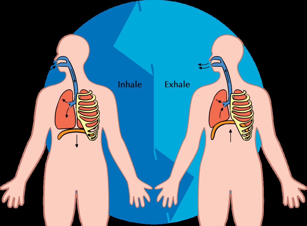
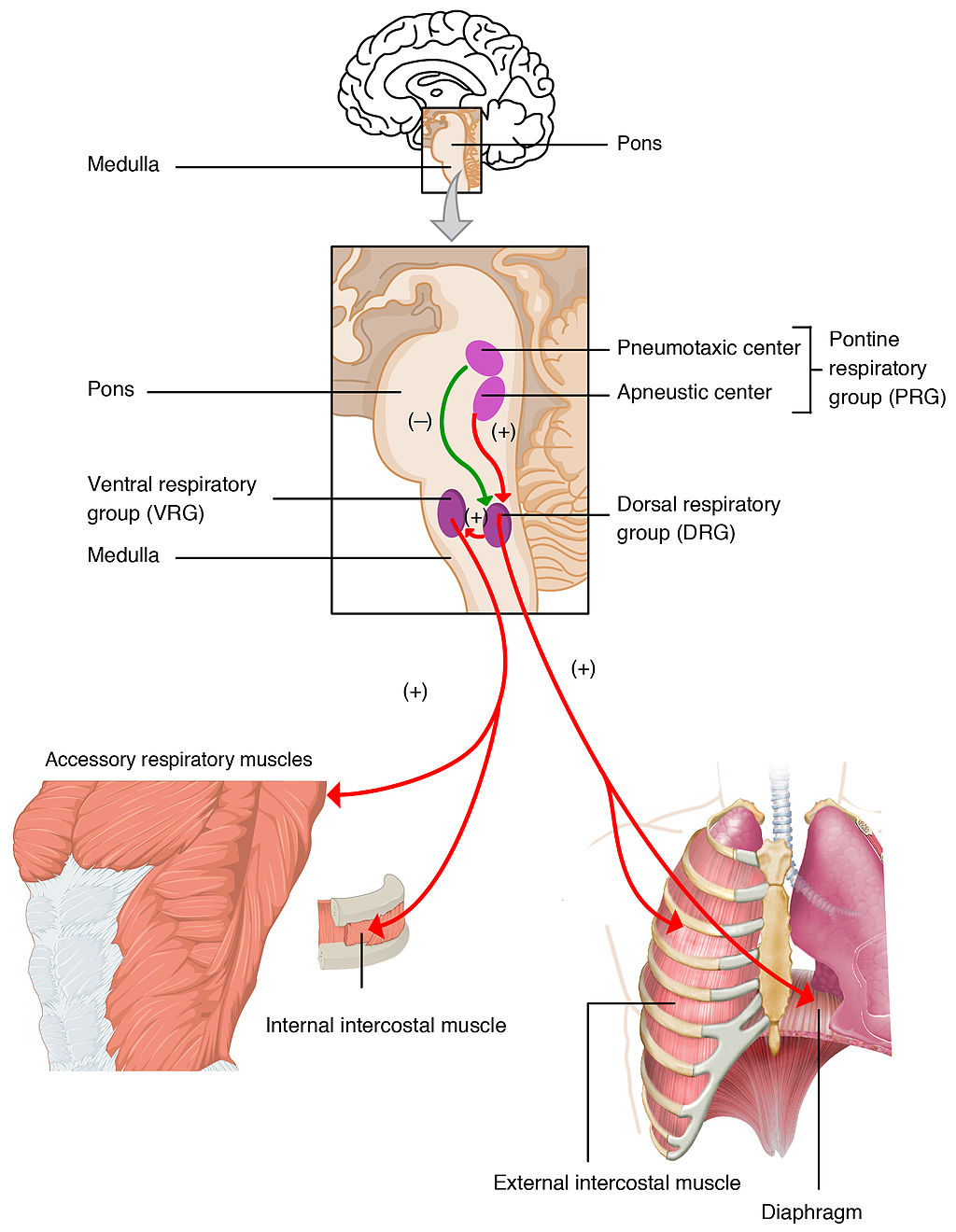
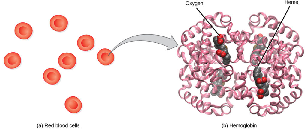
Figure 12.18 Transport of carbon dioxide

Review

  1. Oxyhemoglobin forms by a chemical reaction between which of the following?
    a. hemoglobin and carbon dioxide
    b. carbonic anhydrase and carbon dioxide
    c. hemoglobin and oxygen
    d. carbonic anhydrase and oxygen
  2. In what form is the majority of carbon dioxide transported in the blood?
    a. Bicarbonate ion
    b. Carbaminohemoglobin
    c. Carbonic acid
    d. Carbonic anhydrase
  3. Describe three ways in which carbon dioxide can be transported.

12.7 SMOKING AND HEALTH

Image of a cigarette
Figure 12.19Smoking kills.

SURE DEATH

This anti-smoking photo (Figure 12.18) clearly makes the point that smoking causes death. The image is not using hyperbole, because smoking actually is deadly. It causes about 7 million deaths each year and is the single greatest cause of preventable death worldwide. As many as half of all people who smoke tobacco die from it. As a result of smoking’s deadly effects, the life expectancy of long-term smokers is significantly less than that of non-smokers. In fact, long-term smokers can expect their lifespan to be reduced by as much as 18 years, and they are three times more likely than non-smokers to die before the age of 70.

WHY IS SMOKING DEADLY?

As shown in Figure 12.20, tobacco smoking has adverse effects on just about every bodily system and organ. The detrimental health effects of smoking depend on the number of years that a person smokes and how much the person smokes. Contrary to popular belief, all forms of tobacco smoke — including smoke from cigars and tobacco pipes — have similar health risks as those of cigarette smoke. Smokeless tobacco may be less of a danger to the lungs and heart, but it, too, has serious health effects. It significantly increases the risk of cancers of the mouth and throat, among other health problems.

15.6.2 Effects of Smoking
Figure 12.20 Smoking is known to cause many different cancers and chronic diseases.

Even non-smokers may not be spared the deadly risks of tobacco smoke. If you spend time around smokers either at home or on the job, then you are at risk of the dangers of secondhand smoke. Secondhand smoke enters the air directly from burning cigarettes (and cigars and pipes), and indirectly from smokers’ lungs. This smoke may linger in indoor air for hours, and it increases the risk of a wide range of adverse health effects. According to Health Canada, second-hand smoke causes 800 deaths from lung cancer and heart disease in non-smokers every year. The 2014 U.S. Surgeon General’s Report concluded that there is no established risk-free level of exposure to secondhand smoke. Non-smokers who are exposed to secondhand smoke may have as much as a 30 per cent increase in their risk of lung cancer and heart disease.

Tobacco contains nicotine, which is a psychoactive drug. Although nicotine in tobacco smoke does not directly cause cancer or most of the other health risks of smoking, it is a highly addictive drug. Nicotine is actually even more addictive than cocaine or heroin. The addictive nature of nicotine explains why it is so difficult for smokers to quit the habit, even when they know the health risks and really want to stop smoking. The good news is that if someone does stop smoking, his or her risks of smoking-related diseases and death soon start to fall. By one year after quitting, the risk of heart disease drops to only half of that of a continuing smoker.

SMOKING AND CANCER

One of the main health risks of smoking is cancer, particular cancer of the lung. Because of the increased risk of lung cancer with smoking, the risk of dying from lung cancer before age 85 is more than 20 times higher for a male smoker than for a male non-smoker. As the rate of smoking increases, so does the rate of lung cancer deaths, although the effects of smoking on lung cancer deaths can take up to 20 years to manifest themselves, as shown in Figure 12.21. Besides lung cancer, several other forms of cancer are also significantly more likely in smokers than non-smokers, including cancers of the kidney, larynx, mouth, lip, tongue, throat, bladder, esophagus, pancreas, and stomach. Unfortunately, many of these cancers have extremely low cure rates.

13.6.3 Smoking vs. Lung Cancer Deaths
Figure 12.21 Cigarette smoking by men in the U.S. began to decline in the 1950s, but it wasn’t until the 1970s — roughly 20 years later — that this was reflected by a concomitant decline in lung cancer deaths in men.

When you consider the composition of tobacco smoke, it’s not surprising that it increases the risk of cancer. Tobacco smoke contains dozens of chemicals proven to be carcinogens or causes of cancer. Many of these chemicals bind to DNA in a smoker’s cells, and may either kill the cells or cause mutations. If the mutations inhibit programmed cell death, the cells can survive to become cancer cells. Some of the most potent carcinogens in tobacco smoke include benzopyrene, acrolein, and nitrosamines. Other carcinogens in tobacco smoke are radioactive isotopes, including lead-210 and polonium-210.

RESPIRATORY EFFECTS OF SMOKING

Long-term exposure to the compounds found in cigarette smoke — such as carbon monoxide and cyanide — are thought to be responsible for much of the lung damage caused by smoking. These chemicals reduce the elasticity of alveoli, leading to chronic obstructive pulmonary disease (COPD). COPD is a permanent, incurable, and often fatal reduction in the capacity of the lungs, reducing the lungs’ ability to fully exhale air. The chronic inflammation that is also present in COPD is exacerbated by the tobacco smoke carcinogen acrolein and its derivatives. COPD is almost completely preventable simply by not smoking and by also avoiding secondhand smoke.

CARDIOVASCULAR EFFECTS OF SMOKING

Inhalation of tobacco smoke causes several immediate responses in the heart and blood vessels. Within one minute of inhalation of smoke, the heart rate begins to rise, increasing by as much as 30 per cent during the first ten minutes of smoking. Carbon monoxide in tobacco smoke binds with hemoglobin in red blood cells, thereby reducing the blood’s ability to carry oxygen. Hemoglobin bound to carbon monoxide forms such a stable complex that it may result in a permanent loss of red blood cell function. Several other chemicals in tobacco smoke lead to narrowing and weakening of blood vessels, as well as an increase in substances that contribute to blood clotting. These changes increase blood pressure and the chances of a blood clot forming and blocking a vessel, thereby elevating the risk of heart attack and stroke. A recent study found that smokers are five times more likely than non-smokers to have a heart attack before the age of 40.

Smoking has also been shown to have a negative impact on levels of blood lipids. Total cholesterol levels tend to be higher in smokers than non-smokers. Ratios of “good” cholesterol to “bad” cholesterol tend to be lower in smokers than non-smokers.

ADDITIONAL ADVERSE HEALTH EFFECTS OF SMOKING

A wide diversity of additional adverse health effects are attributable to smoking. Here are just a few of them:

  • Smokers are at significantly increased risk of developing chronic kidney disease (in addition to kidney cancer). For example, smoking hastens the progression of kidney damage in people with diabetes.
  • People who smoke — especially the elderly — have a greater risk of influenza and other infectious diseases than non-smokers. Smoking more than 20 cigarettes a day has been found to increase the risk of infectious diseases by as much as four times the risk in non-smokers. These effects occur because of damage to both the respiratory system and the immune system.
  • In addition to oral cancer, smoking causes other oral problems, including periodontitis (gum disease). Roughly half of the cases of gum inflammation are attributable to current or former smoking. This inflammation increases the risk of tooth loss, which is also higher in smokers than non-smokers. In addition, smoking stains the teeth and causes halitosis (bad breath).
  • Smoking is a key cause of erectile dysfunction (ED), probably because it leads to narrowing of arteries in the penis, as it does elsewhere in the body. The incidence of ED is about 85 per cent higher in males who smoke than it is in non-smokers.
  • Smoking also has adverse effects on the female reproductive system, potentially causing infertility, in part because it interferes with the body’s ability to produce estrogen. Female smokers are about 60 per cent more likely to be infertile than non-smokers. Pregnant women who smoke or are exposed to secondhand smoke have a higher risk of miscarriages and low-birth-weight infants.
  • Certain therapeutic drugs, including some antidepressants and anticonvulsants, are less effective in smokers than in non-smokers. This occurs because smoking increases levels of liver enzymes that break down the drugs.
  • Smoking causes an estimated ten per cent of all fire-related deaths worldwide. Smokers are also at a greater risk of dying in motor vehicle crashes and other accidents.
  • Smoking leads to an increased risk of bone fractures, especially of the hip. It also leads to slower wound healing after surgery, and an increased rate of postoperative complications.

FEATURE: HUMAN BIOLOGY IN THE NEWS

13.6.4 E-Cigarette
Figure 12.22 An E-Cigarette or Vape.

The item in Figure 12.22 looks like a regular cigarette, but it’s actually an electronic cigarette, or e-cigarette. E-cigarettes are battery-powered devices that change flavored liquids and nicotine into vapor that the user inhales. E-cigarettes are often promoted as being safer than traditional tobacco products, and their use is touted as a good way to quit smoking. They are often not banned in smoke-free areas, where it is illegal to smoke tobacco cigarettes.

A study completed in 2015 by researchers at the Harvard School of Public Health and widely reported in the mass media found that e-cigarettes may, in fact, be very harmful to the user’s health. E-cigarettes contain nicotine and cancer-causing chemicals, such as formaldehyde. According to the study, about three-quarters of flavored e-cigarettes also contain a chemical called diacetyl that causes an incurable and potentially fatal disorder of the lungs, commonly called “popcorn lung” (bronchiolitis obliterans). In this disorder, the bronchioles compress and narrow due to the formation of scar tissue, greatly diminishing the breathing capacity of people with the disorder. Popcorn lung gained its common name in 2004, when it was diagnosed in workers at popcorn factories. The buttery flavoring used in the factories contained diacetyl.

Some manufacturers of e-cigarettes and flavorings advertise that their products are now free of diacetyl. However, because e-cigarettes are not currently regulated by the FDA, there is no way of knowing for sure whether the products are actually safe. Equally disturbing is the appeal of flavored e-cigarettes to teens and producers’ attempts to specifically market their products to this age group. Flavors such as “cotton candy,” “Katy Perry’s cherry,” and “alien blood” are obviously marketed to youth. Not surprisingly, the use of e-cigarettes is on the rise in middle and high school students, who are more likely to use them than regular cigarettes. Public health officials fear that e-cigarettes will be a gateway for teens to move on to smoking tobacco cigarettes. Some U.S. states have recently passed laws prohibiting minors from buying e-cigarettes, and Brazil, Singapore, Uruguay, and India have banned e-cigarettes.

Review

  1. What smoking-related factors determine how smoking affects a smoker’s health?
  2. What are the two sources of secondhand cigarette smoke? How does exposure to secondhand smoke affect non-smokers?
  3. Why is it so difficult for smokers to quit the habit? How is their health likely to be affected by quitting?
  4. Why does smoking cause cancer? List five types of cancer that are significantly more likely in smokers than non-smokers.
  5. Explain how smoking causes COPD.
  6. Do you think e-cigarettes can be addictive? Explain your reasoning.

What you should know about vaping and e-cigarettes, TEDMED, 2019.

12.8 DISORDERS OF THE RESPIRATORY SYSTEM

13.5.1 Dust Mite
Figure 12.23 This is “mitey” scary looking.

A “MITEY” MONSTER

The scary beast in Figure 12.23 is likely to be lurking in your own home, where it feeds on organic debris, including human skin. What is it? It’s the common dust mite, a close relative of spiders. The dust mite is so small that it is barely visible with the

s the common dust mite, a close relative of spiders. The dust mite is so small that it is barely visible with the unaided eye, so it’s obviously shown greatly enlarged above. If you think you can get rid of dust mites in your home by frequent and thorough cleaning, think again. There may be thousands of dust mites in just one gram of dust! Regardless of how clean you keep your house, you can’t eliminate dust mites entirely. So why even bother trying? The feces of dust mites contain proteins that are a common trigger of asthma attacks.

ASTHMA


13.5.2 Asthma
Figure 12.24 During an asthma attack, airways narrow and may become clogged with mucus, making breathing difficult.

Asthma is a chronic inflammatory disease of the airways in the lungs, in which the airways periodically become inflamed. As you can see in Figure 12.24, this causes swelling and narrowing of the airways, often accompanied by excessive mucus production. Symptoms of asthma include difficulty breathing, coughing, wheezing, shortness of breath, and chest tightness. Some people with asthma rarely experience symptoms, and then usually only in response to certain triggers in the environment. Other people may have symptoms almost all of the time.

Asthma is thought to be caused by a combination of genetic and environmental factors. A person with a family history of asthma is more likely to develop the disease. Dozens of genes have been found to be associated with asthma, many of which are related to the immune system. Additional risk factors include obesity and sleep apnea. Environmental factors trigger asthma attacks in people who have a genetic predisposition to the disease. Besides dust mite feces, triggers may include other allergens (such as pet dander, cockroaches, and mold), certain medications including aspirin, air pollution, and stress. Symptoms tend to be worse at night and early in the morning. They may also worsen during upper respiratory tract infections, strenuous exercise, or when the airways are exposed to cold air.

13.5.3 Asthma Inhaler
Figure 12.25 Use of inhaled bronchodilators (medications which cause the bronchi to expand) can help patients manage the long-term effects of living with asthma.

There is no cure for asthma at present, but the symptoms of asthma attacks usually can be reversed with the use of inhaled medications called bronchodilators (as shown in Figure 12.25). These medications soothe the constricted air passages and help to re-expand them, making breathing easier. The medications usually start to take effect almost immediately. Other medications can be taken for long-term control of the disease. These medications help prevent asthma attacks from occurring. Corticosteroids are generally considered the most effective treatment for long-term control. Another way to prevent asthma attacks is by avoiding triggers whenever possible.

PNEUMONIA

Another common inflammatory disease of the respiratory tract is pneumonia. In pneumonia, inflammation affects primarily the alveoli, which are the tiny air sacs of the lungs. Inflammation causes some of the alveoli to become filled with fluid so that gas exchange cannot occur, as you can see illustrated in Figure 12.26. Symptoms of pneumonia typically include coughing, chest pain, difficulty breathing, and fever.

13.5.4 Pneumonia
Figure 12.26Fluid-filled alveoli characteristic of pneumonia inhibit normal gas exchange with the blood.

Pneumonia often develops as a consequence of an upper respiratory tract infection (such as the common cold or flu), especially in the very young and the elderly. It is usually caused by bacteria or viruses, although some cases may be caused by other microorganisms, such as fungi. The majority of cases are caused by just a few pathogens, the most common being the bacterium Streptococcus pneumoniae. Pneumonia is more likely to develop in people who have other lung diseases, such as asthma, a history of smoking, heart failure, or a weakened immune system.

Vaccines are available to prevent certain types of bacterial and viral pneumonia, including pneumonia caused by Streptococcus pneumoniae. Treatment of pneumonia depends on the cause. For example, if the disease is caused by bacteria, antibiotics are generally prescribed. In cases of severe pneumonia, hospitalization and supplemental oxygen may be required.

CHRONIC OBSTRUCTIVE PULMONARY DISEASE

Chronic obstructive pulmonary disease (COPD) is a lung disease characterized by chronic poor airflow due to increasing in-elasticity of lung tissue and breakdown of the walls of the alveoli. The main symptoms include shortness of breath and a cough that produces phlegm. These symptoms are usually present for a long period of time, and typically become worse over time. Eventually, walking up stairs and similar activities become difficult because of shortness of breath.

COPD formerly was referred to as chronic bronchitis or emphysema. Now, the term chronic bronchitis is used to refer to the symptoms of COPD, and the term emphysema is used to refer to the lung changes that occur with COPD. Some of these lung changes are shown in Figure 12.27 below. They include a breakdown of connective tissues that reduces the number and elasticity of alveoli. As a result, the patient can no longer fully exhale air from the lungs, so air becomes trapped in the lungs. Gas exchange is hampered and may lead to low oxygen levels, as well as too much carbon dioxide in the blood.

13.5.5 COPD
Figure 12.27 The physiological changes that occur with COPD include a breakdown of alveolar walls, reducing the surface area for gas exchange.

Smoking tobacco and vaping are the major cause of COPD, with a number of other factors such as air pollution and genetics playing smaller roles. Of people who are life-long smokers, about half will eventually develop COPD. Exposure to secondhand smoke in nonsmokers also increases the risk of COPD, and accounts for about 20 per cent of cases.  Most cases of COPD could have been prevented by never smoking or vaping. In people who have already been diagnosed with COPD, cessation of smoking or vaping can slow down the rate at which COPD worsens. People with COPD may be treated with supplemental oxygen and inhaled bronchodilators. These treatments may reduce the symptoms, but there is no cure for COPD except — in very severe cases — lung transplantation.

LUNG CANCER

Lung cancer is a malignant tumor characterized by uncontrolled cell growth in tissues of the lung. The tumor may arise directly from lung tissue (primary lung cancer), or as a result of metastasis from cancer in another part of the body (secondary lung cancer). Primary lung cancer may also metastasize and spread to other parts of the body. Lung cancer develops after genetic damage to DNA that affects the normal functions of the cell. As more damage accumulates, the risk of cancer increases. The most common symptoms of lung cancer include coughing (especially coughing up blood), wheezing, shortness of breath, chest pain, and weight loss.

The major cause of primary lung cancer is tobacco use, which accounts for about 85 per cent of cases. Cigarette smoke contains numerous cancer-causing chemicals. Besides smoking, other potential causes of lung cancer include exposure to radon gas, asbestos, secondhand smoke, or other air pollutants. When tobacco smoking is combined with another risk factor (such as exposure to radon or asbestos), the risk of lung cancer is heightened. People who have close biological relatives with lung cancer are also at increased risk of developing the disease.

Most cases of lung cancer cannot be cured. In many people, the cancer has already spread beyond the original site by the time they have symptoms and seek medical attention. About ten per cent of people with lung cancer do not have symptoms when they are diagnosed, and the cancers are found when they have a chest X-ray for another problem. In part because of its typically late diagnosis, lung cancer is the most common cause of cancer-related death in men, and the second most common cause in women (after breast cancer). Approximately 21,000 Canadians die from lung cancer each year.  Common treatments for lung cancer include surgical removal of the tumor, radiation therapy, chemotherapy, or some combination of these three types of treatment.

FEATURE: MY HUMAN BODY

Do you — or someone you love — snore? Snoring may be more than just an annoyance. It may also be a sign of a potentially dangerous and common disorder known as sleep apnea. Sleep apnea is characterized by pauses in breathing that occur most often because of physical blockage to airflow during sleep. When breathing is paused, carbon dioxide builds up in the bloodstream. The higher-than-normal level of carbon dioxide in the blood causes the respiratory centers in the brain to wake the person enough to start breathing normally. This reduces the carbon dioxide level, and the person falls back asleep. This occurs repeatedly throughout the night, causing serious disruption in sleep. Most people with sleep apnea are unaware that they have the disorder, because they don’t wake up fully enough to remember the repeated awakenings throughout the night. Instead, sleep apnea is more commonly recognized by other people who witness the episodes.

Figure 12.28 below shows how sleep apnea typically occurs. The muscle tone of the body normally relaxes during sleep, allowing the soft tissues in the throat to collapse and block the airway. The relaxation of muscles may be exacerbated by the use of alcohol, tranquilizers, or muscle relaxants. The risk of sleep apnea is greater in people who are overweight, smoke tobacco, or have diabetes. The disorder is also more likely to occur in older people and males. Common symptoms of sleep apnea include loud snoring, restless sleep, and daytime sleepiness and fatigue. Daytime sleepiness, in turn, increases the risk of driving and work-related accidents. Continued sleep deprivation may cause moodiness and belligerence. Lack of adequate oxygen to the body because of sleep apnea may also lead to other health problems, including fatty liver diseases and high blood pressure. Symptoms of sleep apnea may be present for years — or even decades — until (and if) a diagnosis is finally made.

15.5.6 Sleep Apnea Airway Blockage
Figure 12.28 Sleeping on one’s back may increase the risk of the airway becoming temporarily blocked during sleep, resulting in sleep apnea.
15.5.7 CPAP to Treat Sleep Apnea
Figure 12.29 The use of a CPAP machine can be used to treat sleep apnea.

Treatment of sleep apnea may include avoiding alcohol, quitting smoking, or losing weight. Elevating the upper body during sleep or sleeping on one’s side may help prevent airway collapse in people with sleep apnea.

Review

  1. How can asthma attacks be prevented? How can symptoms of asthma attacks be controlled?.
  2. How can pneumonia be prevented? How is it treated?
  3. What is the difference between primary and secondary lung cancer? What is the major cause of primary lung cancer? Discuss lung cancer as a cause of death. How is lung cancer treated?
  4. What is the difference between how COPD and pneumonia affect the alveoli?

CASE STUDY: COUGH THAT WON’T QUIT

Three weeks ago, 20-year-old Erica came down with symptoms typical of the common cold. She had a runny nose, fatigue, and a mild cough. Her symptoms were starting to improve, but recently, her cough has been getting worse. She is coughing up a lot of thick mucus, her throat is sore from frequent coughing, and her chest feels very congested. According to her grandmother, Erica has a “chest cold.” Erica is a smoker and wonders if her habit is making her cough worse. She decides that it’s time to see a doctor.

Dr. Choo examines Erica and asks about her symptoms and health history. She checks the level of oxygen in Erica’s blood by attaching a device called a pulse oximeter to Erica’s finger.

13.1.2 Oximeter
Figure 12.30 A pulse oximeter, used to measure blood oxygen levels.

Dr. Choo concludes that Erica has bronchitis, which is an infection that commonly occurs after a person has a cold or flu. Bronchitis is sometimes referred to as a “chest cold,” so Erica’s grandmother was right! Bronchitis causes inflammation and a build up of mucus in the bronchial tubes in the chest.

Because bronchitis is usually caused by viruses and not bacteria, Dr. Choo tells Erica that antibiotics are not likely to help. Instead, she recommends that Erica try to thin out and remove the mucus by drinking plenty of fluids and using a humidifier or spending time in a steamy shower. She recommends that Erica get plenty of rest as well.

Dr. Choo also tells Erica some things not to do — most importantly, to stop smoking while she is sick, and to try to quit smoking in the long-term. She explains that smoking can make people more susceptible to bronchitis and can hinder recovery. Finally, she advises Erica to avoid taking over-the-counter cough suppressant medication.

Inhaling the moist air from a humidifier or steamy shower can feel particularly good if you have a respiratory system infection, such as bronchitis. The moist air helps to loosen and thin mucus in the respiratory system, allowing you to breathe easier.

In the beginning of this chapter, you learned about Erica, who developed acute bronchitis after getting a cold. She had a worsening cough, a sore throat due to coughing, and chest congestion. She was also coughing up thick mucus.

13.7.2 Bronchitis
Figure 12.31 The function of mucus is to trap pathogens and other potentially dangerous particles that enter the respiratory system from the air. However, when too much mucus is produced in response to an infection (as in the case of bronchitis), it can interfere with normal airflow. The body responds by coughing as it tries to rid itself of the pathogen-laden mucus.

Acute bronchitis usually occurs after a cold or flu, usually due to the same viruses that cause cold or flu. Because bronchitis is not usually caused by bacteria (although it can be), in most cases, antibiotics are not an effective treatment.

Bronchitis affects the bronchial tubes, which, as you have learned, are air passages in the lower respiratory tract. The main bronchi branch off of the trachea and then branch into smaller bronchi, and then bronchioles. In bronchitis, the walls of the bronchi become inflamed, which makes them narrower. There is also excessive production of mucus in the bronchi, which further narrows the pathway where air can flow through. Figure 12.31, shows how bronchitis affects the bronchial tubes.

The treatment for most cases of bronchitis involves thinning and loosening the mucus so that it can be effectively coughed out of the airways. This can be done by drinking plenty of fluids, using humidifiers or steam, and — in some cases — using over-the-counter medications (such as expectorants). Dr. Choo recommended some of these treatments to Erica, and also warned against using cough suppressants. Cough suppressants work on the nervous system to suppress the cough reflex. When a patient has a “productive” cough (which means they are coughing up mucus), doctors generally advise them not to take cough suppressants, so that they can cough the mucus out of their bodies.

When Dr. Choo was examining Erica, she used a pulse oximeter to measure the oxygen level in her blood. Why did she do this? As you have learned, the bronchial tubes branch into bronchioles, which ultimately branch into the alveoli of the lungs. The alveoli are where gas exchange occurs between the air and the blood to take in oxygen and remove carbon dioxide and other wastes. By checking Erica’s blood oxygen level, Dr. Choo was making sure that her clogged airways were not impacting her level of much-needed oxygen.

Erica has acute bronchitis, but you may recall that chronic bronchitis was discussed earlier in this chapter (Section 13.5) as a term that describes the symptoms of chronic obstructive pulmonary disease (COPD). COPD is often due to tobacco smoking, and it causes damage to the walls of the alveoli. Acute bronchitis, on the other hand, typically occurs after a cold or flu, and involves inflammation and mucus build-up in the bronchial tubes. As implied by the difference in their names, chronic bronchitis is an ongoing, long-term condition, while acute bronchitis is likely to resolve relatively quickly with proper rest and treatment.

Erica uses e-cigarettes (vaping), so she is more likely to develop chronic respiratory conditions, such as COPD. As you have learned, smoking damages the respiratory system, along with many other systems of the body. Smoking and vaping increases the risk of respiratory infections, including bronchitis and flu, due to its damaging effects on the respiratory and immune systems. Dr. Choo strongly encouraged Erica to quit vaping, not only so that her acute bronchitis resolves, but so that she can avoid future infections and other negative health outcomes associated with vaping and smoking, including COPD and lung cancer.

As you have learned in this chapter, the respiratory system is critical to carry out the gas exchange necessary for life’s functions, and to protect the body from pathogens and other potentially harmful substances in the air. But this ability to interface with the outside air has a cost. The respiratory system is prone to infections, as well as damage and other negative effects from allergens, mold, air pollution, cigarette smoke and vaping. While exposure to most of these things cannot be avoided, not smoking is an important step you can take to protect this organ system — as well as many other systems of your body.


Attributions

This chapter is composed of text taken from of the following sources:

Cushwa, W., & Senior Contributors. (2015). Human biology. OpenStax CNX. [CC BY 4.0 license]. [Retrieved from Human Biology : Willy Cushwa : Free Download, Borrow, and Streaming : Internet Archive]

Figure 12.17: Transport of carbon dioxide in the blood – Methods of transport. [Image]. Bing Images. Retrieved from [transport of carbon diooxide in blood oer images – Search (bing.com)]

ISKME. (2007-2024). Gas exchange across respiratory surfaces. In Biology, Animal Structure and Function: The Respiratory System. Rice University. OER Commons. [Creative Commons Attribution-NonCommercial-ShareAlike 4.0 License]. Retrieved from [Biology, Animal Structure and Function, The Respiratory System, Gas Exchange across Respiratory Surfaces | OER Commons]

LibreTexts. (n.d.). Figure 12.16: Transport of oxygen in the blood. In Biology. Retrieved from 22.16: Transport of Oxygen in the Blood – Biology LibreTexts

Miller, C. (2020). Human biology: Human anatomy and physiology. Thompson Rivers University. [CC BY NC]. Retrieved from [Human Biology – Simple Book Publishing (tru.ca)]


License

Icon for the Creative Commons Attribution-NonCommercial 4.0 International License

Human Biology Copyright © by Viveca Sulich is licensed under a Creative Commons Attribution-NonCommercial 4.0 International License, except where otherwise noted.

Share This Book