Want to create or adapt books like this? Learn more about how Pressbooks supports open publishing practices.
3 Cells
3.1 Chapter Overview
This chapter will describe cells, which are the basic unit of structure and function in all living organisms. Specifically, you will learn:
How cells were discovered, their common structures, and the principles of cell theory.
The importance of size and shape to the functions of cells.
The differences between eukaryotic cells (such as those in humans and other animals) and prokaryotic cells (such as bacteria).
The structures and functions of cell parts, including mitochondria, the plasma membrane, cytoplasm, cytoskeleton, nucleus, ribosomes, Golgi apparatus, endoplasmic reticulum, vesicles, and vacuoles.
The processes of passive and active transport to move substances into and out of cells and help maintain homeostasis.
How organisms obtain the energy needed for life, including how the sugar glucose is broken down to produce ATP through the processes of anaerobic and aerobic cellular respiration.
The phases of the cell cycle, how cells divide through mitosis, and how cancer can result from unregulated cell division.
3.2 DISCOVERY OF CELLS AND CELL THEORY
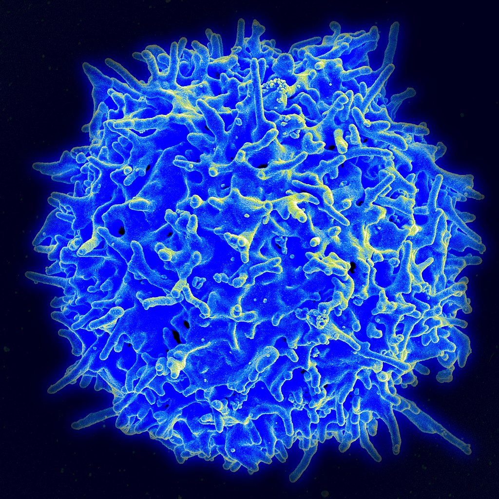
Figure 3.1 Human cells viewed with a very powerful tool called a scanning electron microscope.
AMAZING CELLS
What are these incredible objects? Would it surprise you to learn that they are all human cells. Cells are actually too small to see with the unaided eye. It is visible here in such detail because it is being viewed with a very powerful tool called a scanning electron microscope. Cells may be small in size, but they are extremely important to life. Like all other living things, you are made of cells. Cells are the basis of life, and without cells, life as we know it would not exist. You will learn more about these amazing building blocks of life in this section.
WHAT ARE CELLS?
If you look at living matter with a microscope — even a simple light microscope — you will see that it consists of cells . Cells are the basic units of the structure and function of living things. They are the smallest units that can carry out the processes of life. All organisms are made up of one or more cells, and all cells have many of the same structures and carry out the same basic life processes. Knowing the structure of cells and the processes they carry out is necessary to an understanding of life itself.
DISCOVERY OF CELLS
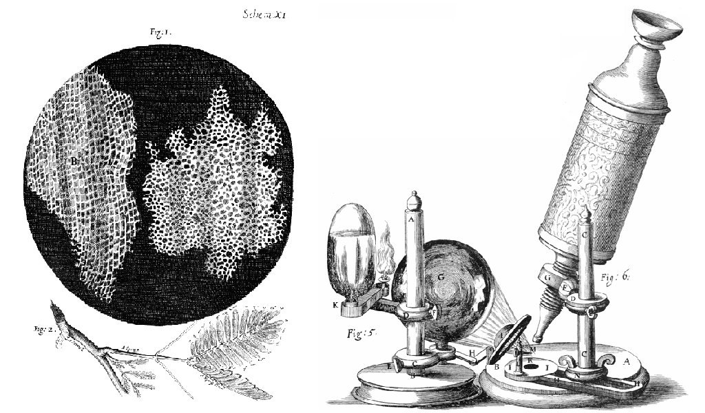
The first time the word cell was used to refer to these tiny units of life was in 1665 by a British scientist named Robert Hooke. Hooke was one of the earliest scientists to study living things under a microscope. The microscopes of his day were not very strong, but Hooke was still able to make an important discovery. When he looked at a thin slice of cork under his microscope, he was surprised to see what looked like a honeycomb. Hooke made the drawing in the figure to the right to show what he saw. As you can see, the cork was made up of many tiny units. Hooke called these units cells because they resembled cells in a monastery.
Figure 3.2 Robert Hooke sketched the cork cells as they appeared under a simple light microscope.
Soon after Robert Hooke discovered cells in cork, Anton van Leeuwenhoek in Holland made other important discoveries using a microscope. Leeuwenhoek made his own microscope lenses, and he was so good at it that his microscope was more powerful than other microscopes of his day. In fact, Leeuwenhoek’s microscope was almost as strong as modern light microscopes. Using his microscope, Leeuwenhoek was the first person to observe human cells and bacteria.
CELL THEORY
By the early 1800s, scientists had observed cells of many different organisms. These observations led two German scientists named Theodor Schwann and Matthias Jakob Schleiden to propose cells as the basic building blocks of all living things. Around 1850, a German doctor named Rudolf Virchow was studying cells under a microscope, when he happened to see them dividing and forming new cells. He realized that living cells produce new cells through division. Based on this realization, Virchow proposed that living cells arise only from other living cells.
The ideas of all three scientists — Schwann, Schleiden, and Virchow — led to cell theory, which is one of the fundamental theories unifying all of biology.
Cell theory states that:
All organisms are made of one or more cells.
All the life functions of organisms occur within cells.
All cells come from existing cells.
SEEING INSIDE CELLS
Starting with Robert Hooke in the 1600s, the microscope opened up an amazing new world — a world of life at the level of the cell. As microscopes continued to improve, more discoveries were made about the cells of living things, but by the late 1800s, light microscopes had reached their limit. Objects much smaller than cells (including the structures inside cells) were too small to be seen with even the strongest light microscope.
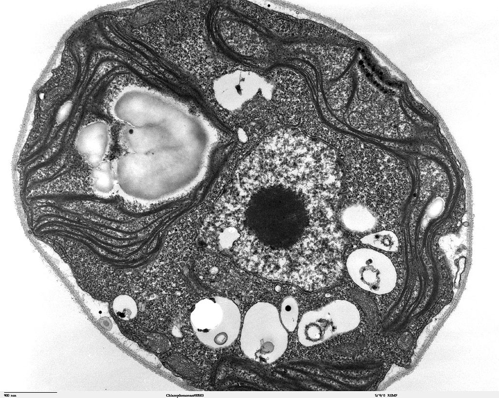
Figure 3.3 An electron microscope produced this image of the structures inside of a cell.
Then, in the 1950s, a new type of microscope was invented. Called the electron microscope, it used a beam of electrons instead of light to observe extremely small objects. With an electron microscope, scientists could finally see the tiny structures inside cells. They could even see individual molecules and atoms. The electron microscope had a huge impact on biology. It allowed scientists to study organisms at the level of their molecules, and it led to the emergence of the molecular biology field. With the electron microscope, many more cell discoveries were made.
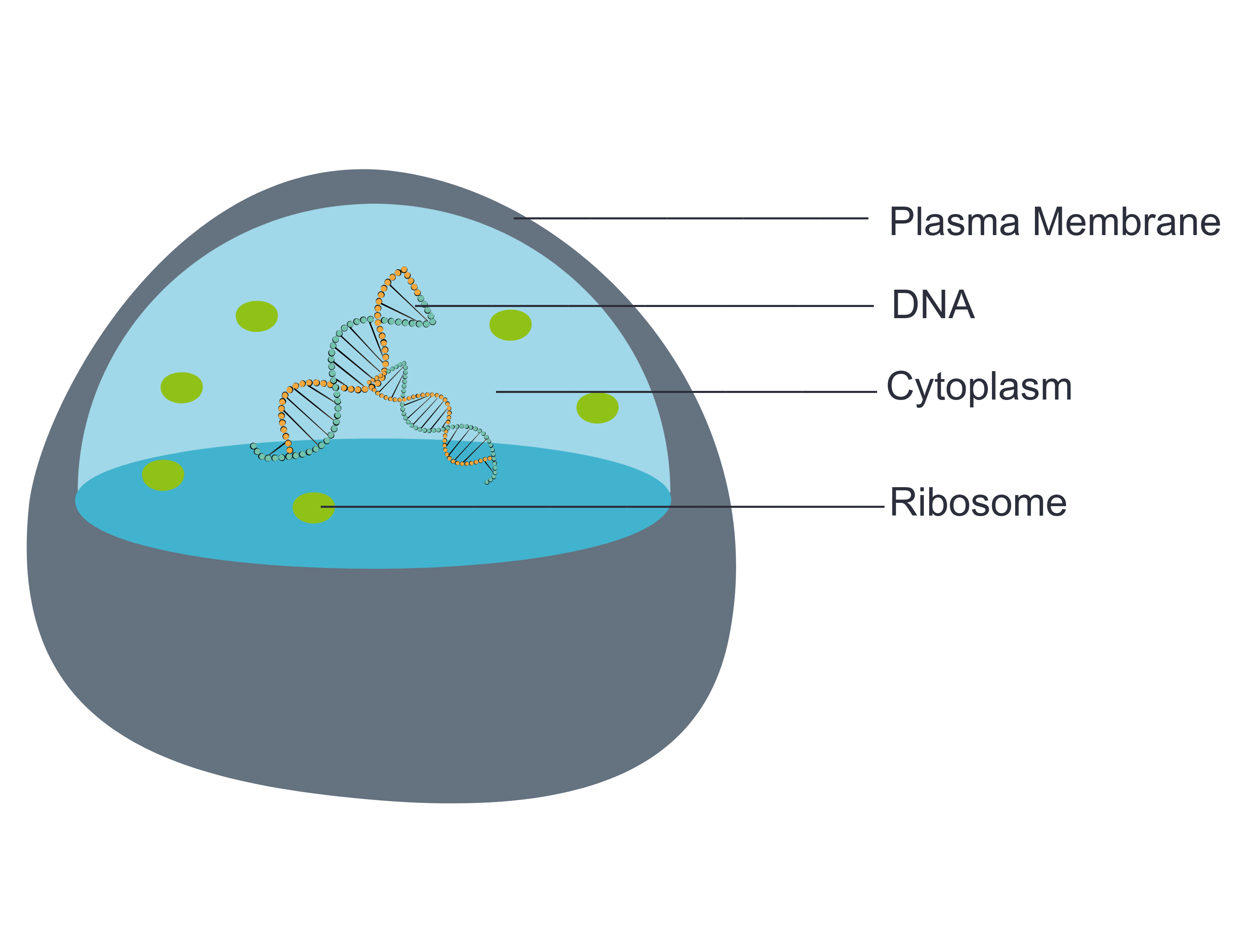
STRUCTURES SHARED BY ALL CELLS
Although cells are diverse, all cells have certain parts in common. These parts include a plasma membrane, cytoplasm, ribosomes, and DNA.
Figure 3.4 Every cell consists of at least a plasma membrane, DNA, ribosomes and a cytoplasm.
The plasma membrane (a type of cell membrane) is a thin coat of lipids that surrounds a cell. It forms the physical boundary between the cell and its environment. You can think of it as the “skin” of the cell.
Cytoplasm refers to all of the cellular material inside of the plasma membrane. Cytoplasm is made up of a watery substance called cytosol, and it contains other cell structures, such as ribosomes.
Ribosomes are the structures in the cytoplasm in which proteins are made.is a nucleic acid found in cells. It contains the genetic instructions that cells need to make proteins.
DNA is a nucleic acid found in cells. It contains the genetic instructions that cells need to make proteins.
These four parts are common to all cells, from organisms as different as bacteria and human beings. How did all known organisms come to have such similar cells? The similarities show that all life on Earth has a common evolutionary history.
Review
Describe cells.
Explain how cells were discovered.
Outline the development of cell theory.
Identify the structures shared by all cells.
The first person to give cells their name based on what he observed in thin slices of cork under the microscope was ____________.
_____________ observed many types of organisms using the microscope and proposed that cells were the basic building blocks of all living things.
Explore More
Watch this video on cells.
3.3 VARIATION IN CELLS
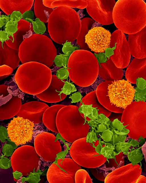
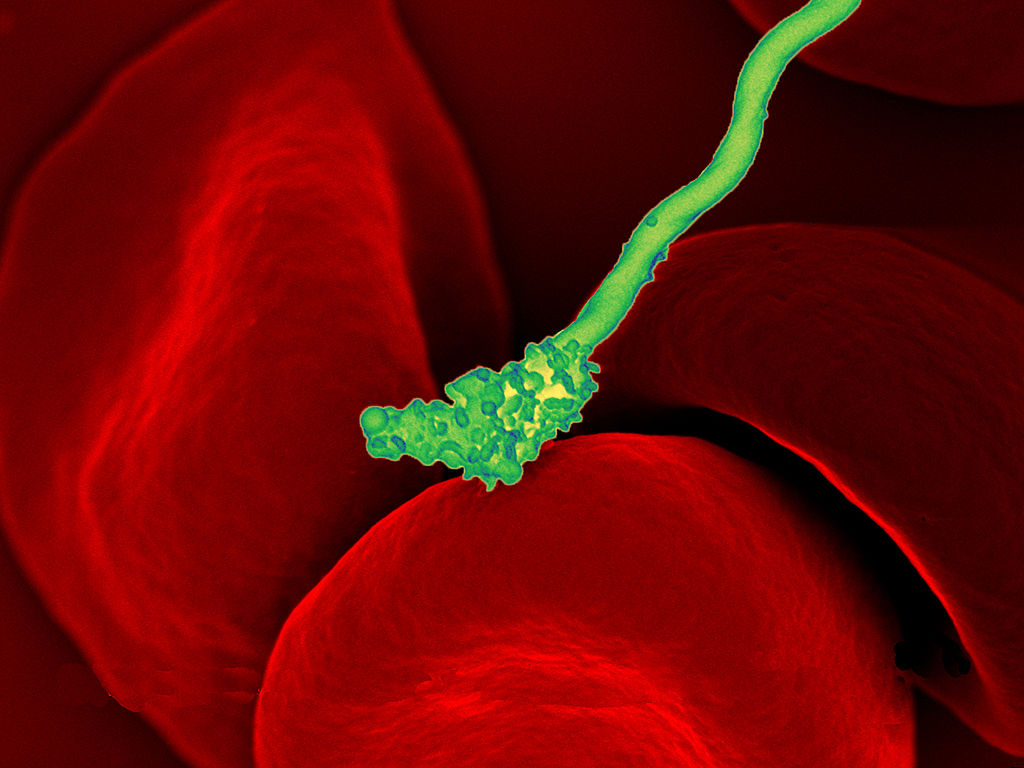
Figure 3.5 A bacterium attacks a human erythrocyte. Both are cells.
BACTERIA ATTACK!
The colorful image in Figure 3.5 shows a bacterial cell (in green) attacking human red blood cells. The bacterium causes a disease called relapsing fever. The bacterial and human cells look very different in size and shape. Although all living cells have certain things in common — such as a plasma membrane and cytoplasm — different types of cells, even within the same organism, may have their own unique structures and functions. Cells with different functions generally have different shapes that suit them for their particular job. Cells vary not only in shape, but also in size, as this example shows. In most organisms, however, even the largest cells are no bigger than the period at the end of this sentence. Why are cells so small?
EXPLAINING CELL SIZE
Most organisms, even very large ones, have microscopic cells. Why don’t cells get bigger instead of remaining tiny and multiplying? Why aren’t you one giant cell rolling around school? What limits cell size?
Once you know how a cell functions, the answers to these questions are clear. To carry out life processes, a cell must be able to quickly pass substances in and out of the cell. For example, it must be able to pass nutrients and oxygen into the cell and waste products out of the cell. Anything that enters or leaves a cell must cross its outer surface. The size of a cell is limited by its need to pass substances across that outer surface. The size of a cell is limited by its need to pass substances across that outer surface.
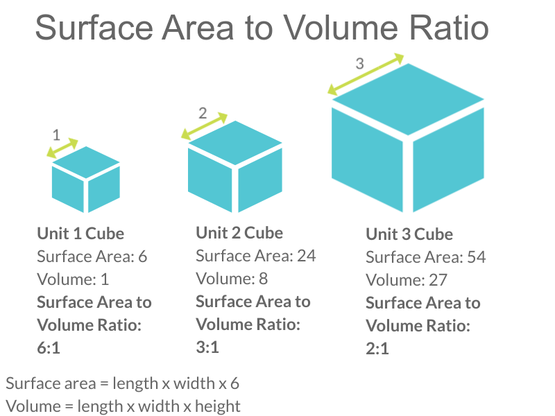
Look at the three cubes in Figure 3.6. A larger cube has less surface area relative to its volume than a smaller cube. This relationship also applies to cells — a larger cell has less surface area relative to its volume than a smaller cell. A cell with a larger volume also needs more nutrients and oxygen and produces more waste. Because all of these substances must pass through the surface of the cell, a cell with a large volume will not have enough surface area to allow it to meet its needs. The larger the cell is, the smaller its ratio of surface area to volume, and the more difficult it will be for the cell to get rid of its waste and take in necessary substances. This is what limits the size of the cell.
Figure 3.6 Surface area to volume ratio.
CELL FORM AND FUNCTION
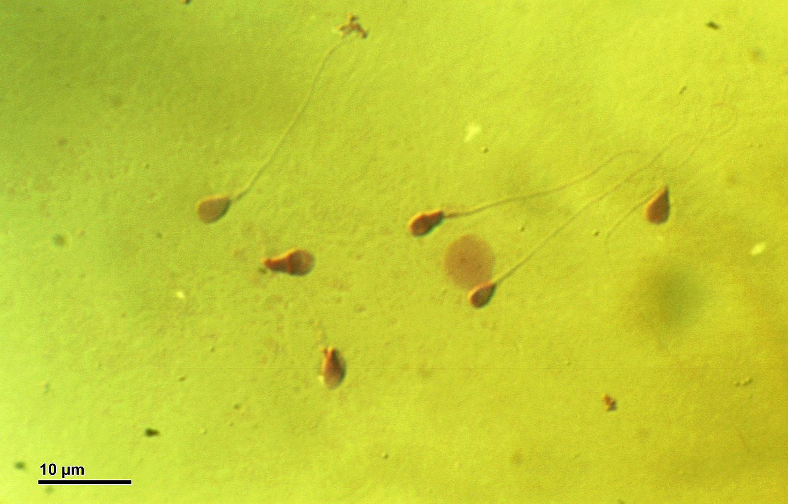
Cells with different functions often have varying shapes. The cells pictured below (Figure 3.7) are just a few examples of the many different shapes that human cells may have. Each type of cell has characteristics that help it do its job. The job of the nerve cell, for example, is to carry messages to other cells. The nerve cell has many long extensions that reach out in all directions, allowing it to pass messages to many other cells at once. Do you see the tail of each tiny sperm cell? Its tail helps a sperm cell “swim” through fluids in the female reproductive tract in order to reach an egg cell. The white blood cell has the job of destroying bacteria and other pathogens. It is a large cell that can engulf foreign invaders.
Figure 3.7 Human cells may have many different shapes that help them to do their jobs.
CELLS WITH AND WITHOUT A NUCLEUS
The nucleus is a basic cell structure present in many — but not all — living cells. The nucleus of a cell is a structure in the cytoplasm that is surrounded by a membrane (the nuclear membrane) and contains DNA. Based on whether or not they have a nucleus, there are two basic types of cells: prokaryotic cells and eukaryotic cells.
PROKARYOTIC CELLS
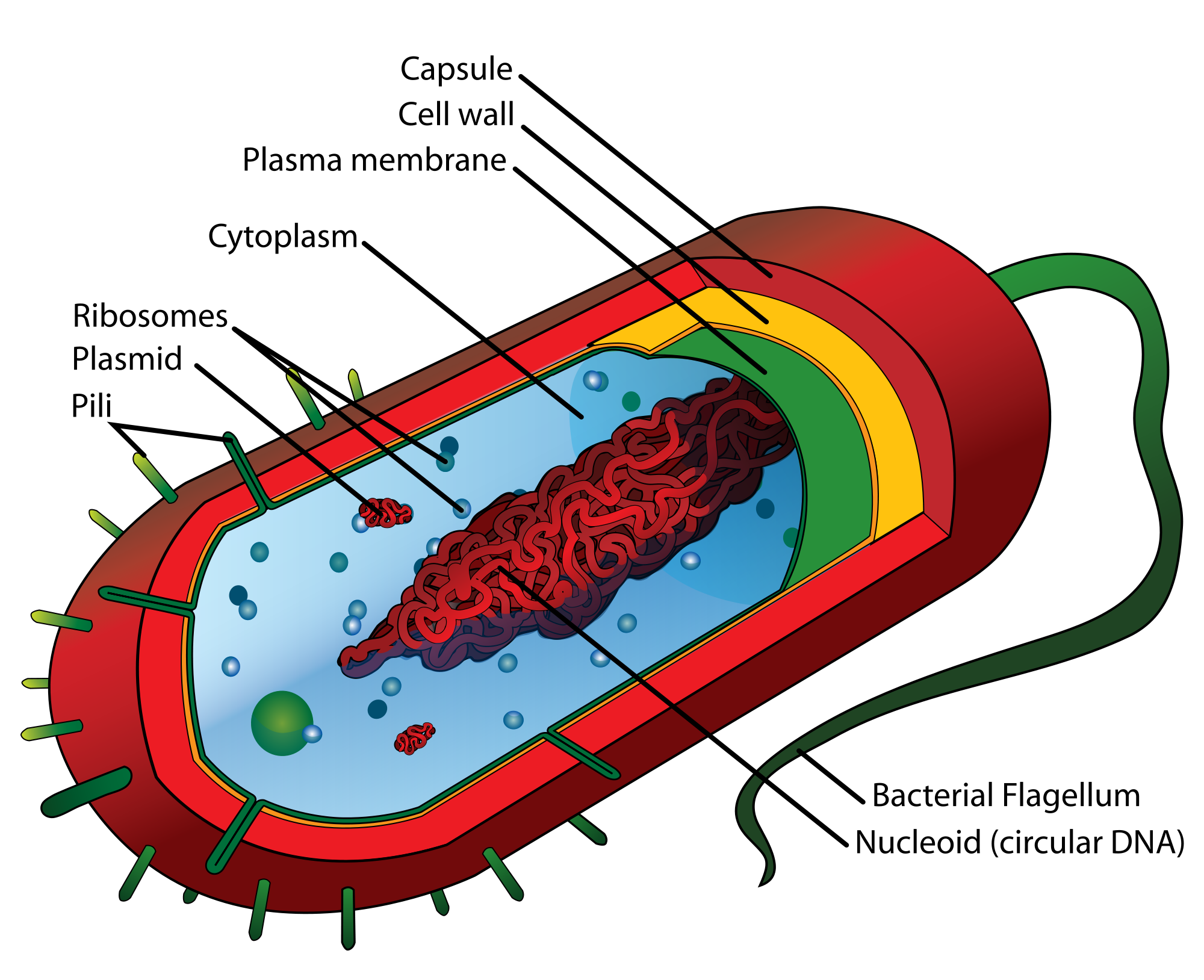
Prokaryotic cells are cells without a nucleus. The DNA in prokaryotic cells is in the cytoplasm, rather than enclosed within a nuclear membrane. In addition, these cells are typically smaller than eukaryotic cells and contain fewer organelles. Prokaryotic cells are found in single-celled organisms, such as the bacterium represented by the model in Figure 3.8. Organisms with prokaryotic cells are called prokaryotes. They were the first type of organisms to evolve, and they are still the most common organisms today.
Figure 3.8 Bacteria are prokaryotes, meaning they do not have a nucleus. Their DNA is contained in a region called the nucleoid.
EUKARYOTIC CELLS
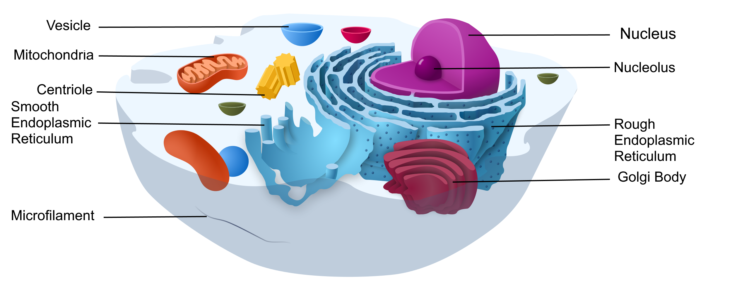
Figure 3.9 Eukaryotic cells, like this animal cell, contain a nucleus and many other membrane-bound organelles.
Eukaryotic cells are cells that contain a nucleus. A typical eukaryotic cell is represented by the model in Figure 3.9. Eukaryotic cells are usually larger than prokaryotic cells. They are found in some single-celled and all multicellular organisms. Organisms with eukaryotic cells are called eukaryotes, and they range from fungi to humans.
Besides a nucleus, eukaryotic cells also contain other organelles. An organelle is a structure within the cytoplasm that performs a specific job in the cell. Organelles called mitochondria, for example, provide energy to the cell, and organelles called vesicles store substances in the cell. Organelles allow eukaryotic cells to carry out more functions than prokaryotic cells can.
Review
Explain why most cells are very small.
Discuss variations in the form and function of cells.
Cells that lack a nucleus are __________________.
Cells that have many membrane-bound organelles are _______________.
True or False. Prokaryotic cells do not have mitochondria.
Do human cells have organelles? Explain your answer.
Which are usually larger – prokaryotic or eukaryotic cells? What do you think this means for their relative ability to take in needed substances and release wastes? Discuss your answer.
DNA in eukaryotes is enclosed within the _______ ________.
Name three different types of cells in humans.
Which organelle provides energy in eukaryotic cells?
3.4 PLASMA MEMBRANE
A BAG FULL OF JELL-O
A cell resembles a plastic bag full of Jell-O. Its basic structure is a plasma membrane filled with cytoplasm. Like Jell-O containing mixed fruit, the cytoplasm of the cell also contains various structures, including a nucleus and other organelles. Your body is composed of trillions of cells, but all of them perform the same basic life functions. They all obtain and use energy, respond to the environment, and reproduce. How do your cells carry out these basic functions and keep themselves — and you — alive? To answer these questions, you need to know more about the structures that make up cells, starting with the plasma membrane.
WHAT IS THE PLASMA MEMBRANE?
The plasma membrane is a structure that forms a barrier between the cytoplasm inside the cell and the environment outside the cell. Without the plasma membrane, there would be no cell. Although it is very thin and flexible, the plasma membrane protects and supports the cell by controlling everything that enters and leaves it. It allows only certain substances to pass through, while keeping others in or out. To understand how the plasma membrane controls what passes into or out of the cell, you need to know its basic structure.
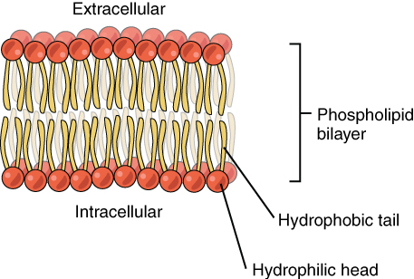
PHOSPHOLIPID BILAYER
The plasma membrane is composed mainly of phospholipids, which consist of fatty acids and alcohol. The phospholipids in the plasma membrane are arranged in two layers, called a phospholipid bilayer. As shown in the simplified diagram in Figure 3.10, each individual phospholipid molecule has a phosphate group head (in red) and two fatty acid tails (in yellow). The head “loves” water (hydrophilic) and the tails “hate” water (hydrophobic). The water-hating tails are on the interior of the membrane, whereas the water-loving heads point outward, toward either the cytoplasm (intracellular) or the fluid that surrounds the cell (extracellular).
One of the great wonders of the cell membrane is its ability to regulate the concentration of substances inside the cell. These substances include ions such as Ca2+, Na+, K+, and Cl–; nutrients including sugars, fatty acids, and amino acids; and waste products, particularly carbon dioxide (CO2), which must leave the cell.
The membrane’s lipid bilayer structure provides the first level of control. The phospholipids are tightly packed, and the membrane has a hydrophobic interior. This structure alone creates what is known as a selectively permeable barrier, one that only allows substances meeting certain physical criteria to pass through it. In the case of the cell membrane, only relatively small, nonpolar materials can move through the lipid bilayer at biologically relevant rates (remember, the lipid tails of the membrane are nonpolar).
Hydrophobic molecules can easily pass through the plasma membrane if they are small enough, because they are water-hating like the interior of the membrane. Hydrophilic molecules, on the other hand, cannot pass through the plasma membrane — at least not without help — because they are water-loving like the exterior of the membrane.
Figure 3.10 The phospholipid bilayer is made up of two sheets of phospholipids, with the fatty acid tails facing the center.
OTHER MOLECULES IN THE PLASMA MEMBRANE
The plasma membrane also contains other molecules, primarily other lipids and proteins. The yellow molecules in the diagram here, for example, are the lipid cholesterol. Molecules of the steroid lipid cholesterol help the plasma membrane keep its shape. Proteins in the plasma membrane (shown blue in Figure 3.11) include: transport proteins that assist other substances in crossing the cell membrane, receptors that allow the cell to respond to chemical signals in its environment, and cell-identity markers that indicate what type of cell it is and whether it belongs in the body.
Figure 3.11 The plasma membrane contains many molecules embedded in the lipid bilayer.
. Other structures:
Protein channels. These span the full membrane and have a space within them because they are used to transport materials into or out of the cell.
Transmembrane proteins. The root “trans” explains that these spans (go “across”) the membrane. Transmembrane proteins can have a variety of functions.
Peripheral proteins. These are found only on one side of the membrane. They can be found on either the cytoplasmic side or the outside of the membrane.
Glycoproteins. These consist of a protein in the plasma membrane with chains of carbohydrates projecting out of the cell.
Glycolipids. These are chains of carbohydrates attached directly to a lipid in the membrane. Both glycoproteins and glycolipids act as labels to identify the cell.
Filaments of cytoskeleton are found along the cytoplasmic side of the membrane and provide a scaffolding for the membrane.
ADDITIONAL FUNCTIONS OF THE PLASMA MEMBRANE
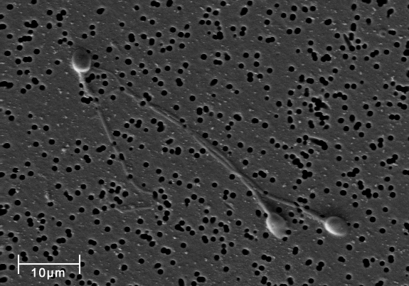
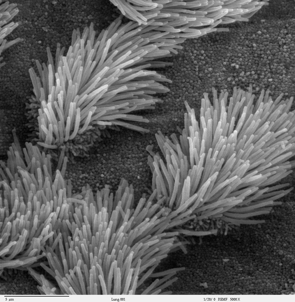
The plasma membrane may have extensions, such as whip-like flagella (singular flagellum) or brush-like cilia (singular cilium), shown below (Figure 3.12), that give it other functions. In single-celled organisms, these membrane extensions may help the organisms move. In multicellular organisms, the extensions have different functions. For example, the cilia on human lung cells sweep foreign particles and mucus toward the mouth and nose, while the flagellum on a human sperm cell allows it to swim.
Figure 3.12 On the left, human sperm with their long, whip-like flagella. On the right, brush-like cilia on lung epithelial cells.
Review
What are the general functions of the plasma membrane?
Describe the phospholipid bilayer of the plasma membrane.
Identify other molecules in the plasma membrane. State their functions.
Why do some cells have plasma membrane extensions, like flagella and cilia?
Explain why hydrophilic molecules cannot easily pass through the cell membrane. What type of molecule in the cell membrane might help hydrophilic molecules pass through it?
Which part of a phospholipid molecule in the plasma membrane is made of fatty acid chains? Is this part hydrophobic or hydrophilic?
The two layers of phospholipids in the plasma membrane are called a phospholipid ____________.
What is the name of the long, whip-like extensions of the plasma membrane that helps some single-celled organisms move?
Why is it important for the plasma membrane to be selectively permeable?
3.5 CYTOPLASM AND CYTOSKELETON
Figure 3.13 The cytoplasm is filled with many organelles, each doing their own specific jobs.
A PEEK INSIDE THE CELL
Figure 3.13 is an artist’s representation of what you might see if you could take a peek inside one of these basic building blocks of living things. A cell’s interior is obviously a crowded and busy space. It contains cytoplasm, dissolved substances, and many structures. It’s a hive of countless biochemical activities all going on at once.
CYTOPLASM
Cytoplasm is a thick, usually colorless solution that fills each cell and is enclosed by the plasma membrane. Cytoplasm presses against the cell membrane, filling out the cell and giving it its shape. Sometimes, cytoplasm acts like a watery solution, and sometimes, it takes on a more gel-like consistency. In eukaryotic cells, the cytoplasm includes all of the material inside the cell but outside of the nucleus which contains its own watery substance called the nucleoplasm. All of the organelles in eukaryotic cells (such as the endoplasmic reticulum and mitochondria) are located in the cytoplasm. The cytoplasm helps to keep them in place. It is also the site of most metabolic activities in the cell, and it allows materials to pass easily throughout the cell.
The portion of the cytoplasm surrounding organelles is called cytosol. Cytosol is the liquid part of cytoplasm. It is composed of about 80 per cent water, and it contains dissolved salts, fatty acids, sugars, amino acids and proteins (such as enzymes). These dissolved substances are needed to keep the cell alive and carry out metabolic processes. Enzymes dissolved in cytosol, for example, break down larger molecules into smaller products that can then be used by organelles of the cell. Waste products are also dissolved in the cytosol before they are taken in by vacuoles or expelled from the cell.
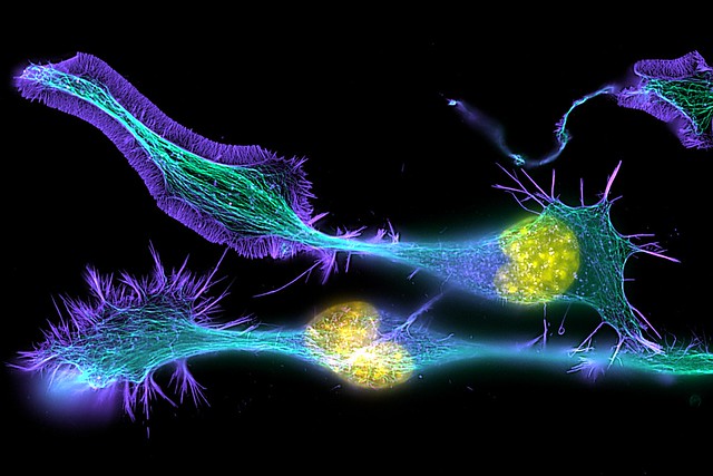
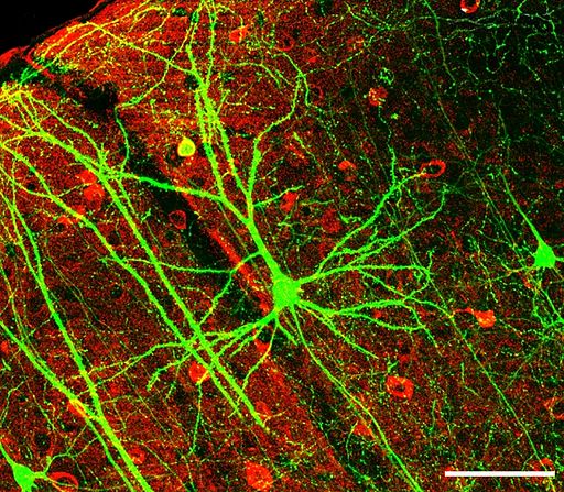
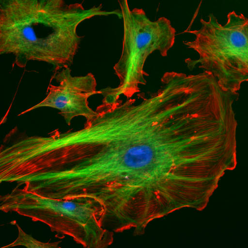
Figure 3.14 The cytoskeleton gives the cell an internal structure, like the frame of a house. In this photograph, filaments and tubules of the cytoskeleton have been stained green and red, respectively, so that they can be seen clearly. The blue dots are cell nuclei.
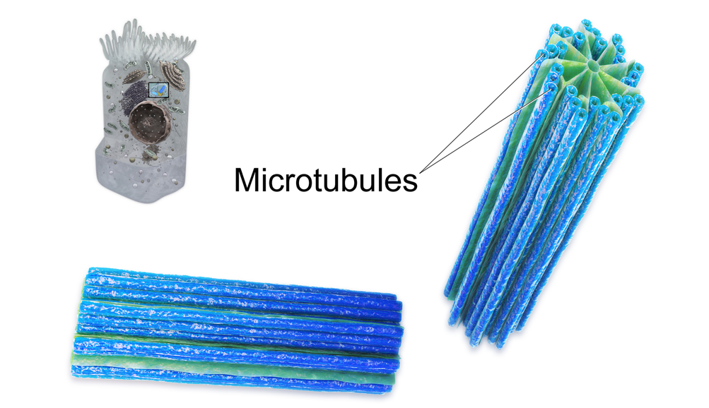
CYTOSKELETON
Although cytoplasm may appear to have no form or structure, it is actually highly organized. A framework of protein scaffolds called the cytoskeleton provides the cytoplasm and the cell with structure. The cytoskeleton consists of thread-like microfilaments, intermediate filaments, and microtubules that crisscross the cytoplasm. As its name suggests, the cytoskeleton is like a cellular “skeleton.” It helps the cell maintain its shape and also helps to hold cell structures (like organelles) in place within the cytoplasm.
FEATURE: HUMAN BIOLOGY IN THE NEWS
News about an important study of the cytoplasm of eukaryotic cells came out in early 2016. Researchers in Dresden, Germany discovered that when cells are deprived of adequate nutrients, they may essentially shut down and become dormant. Specifically, when cells do not get enough nutrients, they shut down their metabolism, their energy level drops, and the pH of their cytoplasm decreases. Their normally liquid cytoplasm also assumes a solid state. The cells appear dead, as though a kind of rigor mortis has set in. The researchers think that these changes protect the sensitive structures inside the cells and allow the cells to survive difficult conditions. If nutrients are returned to the cells, they can emerge from their dormant state unharmed. They will continue to grow and multiply when conditions improve.
This important basic science research was executed on a nonhuman organism: one-celled fungi called yeasts. Nonetheless, it may have important implications for humans, because yeasts have eukaryotic cells with many of the same structures as human cells. Yeast cells appear to be able to “trick” death by shutting down all life processes in a controlled way. Through continued study, researchers hope to learn whether human cells can be taught this “trick” as well.
Review
Describe the composition of cytoplasm. Draw a picture of a cell, including the basic components required to be considered a cell, and the organelles you have learned about in this section.
What are some of the functions of cytoplasm?
Outline the structure and functions of the cytoskeleton.
Is the cytoplasm made of cells? Why or why not?
Name two types of cytoskeletal structures.
Describe one example of a metabolic process that happens in the cytosol.
In eukaryotic cells, all of the material inside of the cell, but outside of the nucleus is called the ___________ .
What is the liquid part of cytoplasm called?
What chemical substance composes most of the cytosol?
Explore more
3.6 CELL ORGANELLES
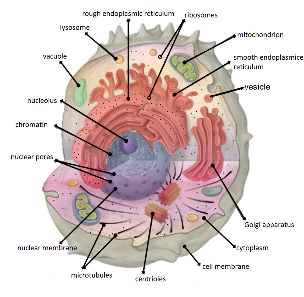
THE NUCLEUS
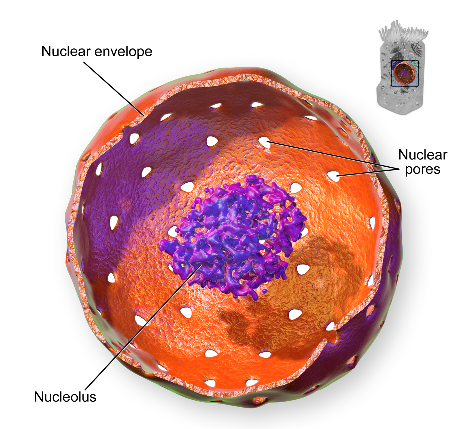
The nucleus the largest organelle in a eukaryotic cell, and it’s considered the cell’s control center. It contains most of the cell’s DNA (which makes up chromosomes), and it is encoded with the genetic instructions for making proteins. The function of the nucleus is to regulate gene expression, including controlling which proteins the cell makes. In addition to DNA, the nucleus contains a thick liquid called nucleoplasm, which is similar in composition to the cytosol found in the cytoplasm outside the nucleus. Most eukaryotic cells contain just a single nucleus, but some types of cells (such as red blood cells) contain no nucleus and a few other types of cells (such as muscle cells) contain multiple nuclei.
As you can see in the model pictured in Figure 3.15, the membrane enclosing the nucleus is called the nuclear envelope. This is actually a double membrane that encloses the entire organelle and isolates its contents from the cellular cytoplasm. Tiny holes called nuclear pores allow large molecules to pass through the nuclear envelope, with the help of special proteins. Large proteins and RNA molecules must be able to pass through the nuclear envelope so proteins can be synthesized in the cytoplasm and the genetic material can be maintained inside the nucleus. The nucleolus shown in the model below is mainly involved in the assembly of ribosomes. After being produced in the nucleolus, ribosomes are exported to the cytoplasm, where they are involved in the synthesis of proteins.
Figure 3.15 This closeup of a cell nucleus shows that it is surrounded by a structure called the nuclear envelope, which contains tiny perforations, or pores. The nucleus also contains a dense center called the nucleolus.
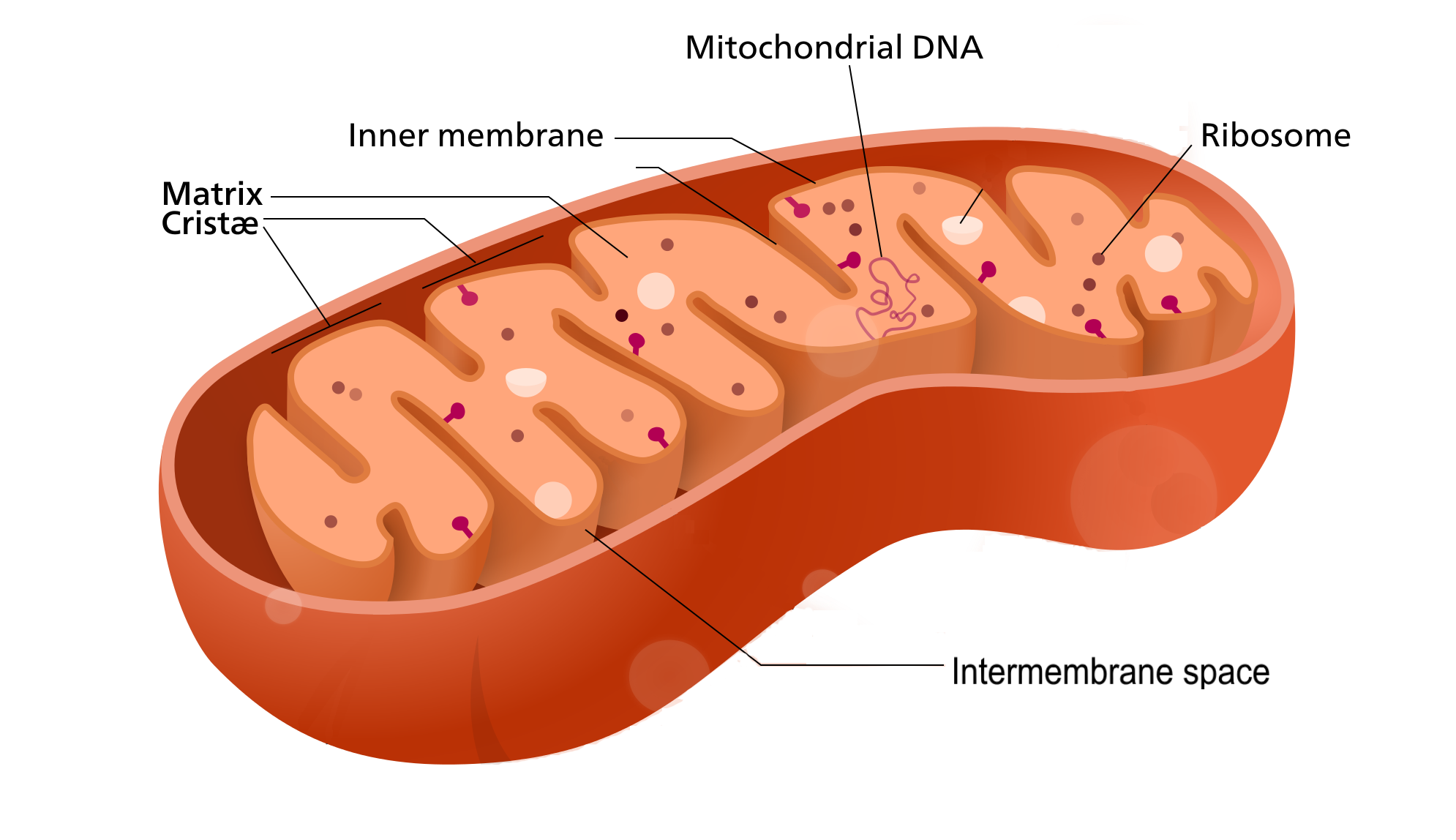
MITOCHONDRIA
The mitochondrion (plural, mitochondria is an organelle that makes energy available to the cell. This is why mitochondria are sometimes referred to as the “power plants of the cell.” They use energy from organic compounds (such as glucose) to make molecules of ATP (adenosine triphosphate) an energy-carrying molecule that is used almost universally inside cells for energy.
Figure 3.16 Mitochondria contain their own DNA and ribosomes!
Mitochondria (as in the Figure 3.16 diagram) have a complex structure including an inner and out membrane. In addition, mitochondria have their own DNA, ribosomes, and a version of cytoplasm, called matrix. Does this sound similar to the requirements to be considered a cell? That’s because they are!
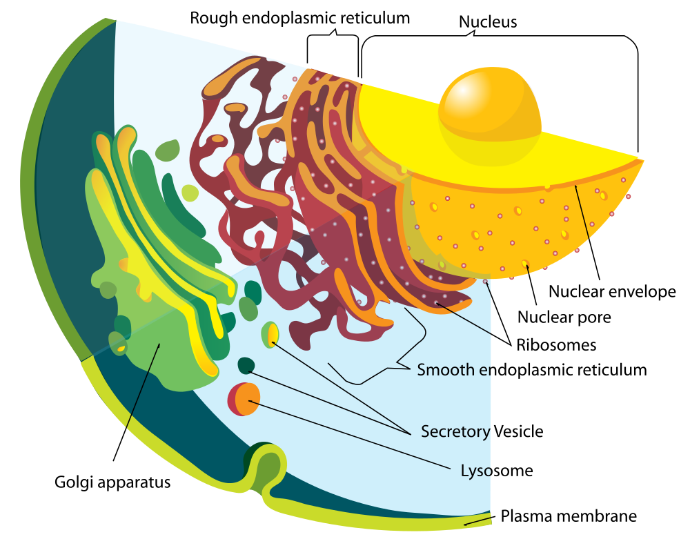
ENDOPLASMIC RETICULUM
The endoplasmic reticulum (ER) is an organelle that helps make and transport proteins and lipids. There are two types of endoplasmic reticulum: rough endoplasmic reticulum (rER) and smooth endoplasmic reticulum (sER). Both types are shown in Figure 3.17.
rER looks rough because it is studded with ribosomes. It provides a framework for the ribosomes, which make proteins. Bits of its membrane pinch off to form tiny sacs called vesicles, which carry proteins away from the ER.
sER looks smooth because it does not have ribosomes. sER makes lipids, stores substances, and plays other roles.
Figure 3.17 The rough and smooth ER are part of a larger group of organelles termed “the endomembrane system”. All of the organelles in this system are composed of plasma membrane.
The Figure 3.17 drawing includes the nucleus, rER, sER, and Golgi apparatus. From the drawing, you can see how all these organelles work together to make and transport proteins.
GOLGI APPARATUS
The Golgi Apparatus (shown in the Figure 3.17 diagram) is a large organelle that processes proteins and prepares them for use both inside and outside the cell. You can see the Golgi apparatus in the figure above. The Golgi apparatus is something like a post office. It receives items (proteins from the ER), then packages and labels them before sending them on to their destinations (to different parts of the cell or to the cell membrane for transport out of the cell). The Golgi apparatus is also involved in the transport of lipids around the cell.
VESICLES AND VACUOLES
Both vesicles and vacuoles are sac-like organelles made of phospholipid bilayer that store and transport materials in the cell. Vesicles are much smaller than vacuoles and have a variety of functions. The vesicles that pinch off from the membranes of the ER and Golgi apparatus store and transport protein and lipid molecules.
There are some vesicles which are specialized to carry out specific functions. Lysosomes, which use enzymes to break down foreign matter and dead cells, have a double membrane to make sure their contents don’t leak into the rest of the cell. Peroxisomes are another type of specialized vesicle with the main function of breaking down fatty acids and some toxins.
CENTRIOLES
Figure 3.18 Centrioles are tiny cylinders near the nucleus, enlarged here to show their tubular structure.
Centrioles are organelles involved in cell division The function of centrioles is to help organize the chromosomes before cell division occurs so that each daughter cell has the correct number of chromosomes after the cell divides. Centrioles are found only in animal cells and are located near the nucleus. Each centriole is made mainly of a protein named tubulin. The centriole is cylindrical in shape and consists of many microtubules, as shown in the model pictured in Figure 3.18.
Figure 3.19 Ribosomes are made up of two subunits, each consisting of protein andrRNA.
RIBOSOMES
Ribosomes are small structures where proteins are made. Although they are not enclosed within a membrane, they are frequently considered organelles. Each ribosome is formed of two subunits.in Figure 3.19. Both subunits consist of proteins and RNA. mRNA from the nucleus carries the genetic code, copied from DNA, which remains in the nucleus. At the ribosome, the genetic code in mRNA is used to assemble and join together amino acids to make proteins. Ribosomes can be found alone or in groups within the cytoplasm, as well as on the rER.
Review
Describe the structure and function of the nucleus.
Explain how the nucleus, ribosomes, rough endoplasmic reticulum, and Golgi apparatus work together to make and transport proteins.
Why are mitochondria referred to as the “power plants of the cell”?
What roles are played by vesicles and vacuoles?
Why do all cells need ribosomes — even prokaryotic cells that lack a nucleus and other cell organelles?
Explore more with these videos
Biology: Cell Structure I Nucleus Medical Media, Nucleus Medical Media, 2015.
David Bolinsky: Visualizing the wonder of a living cell, TED, 2007.
3.7 TRANSPORT ACROSS MEMBRANES
If a cell were a house, the plasma membrane would be walls with windows and doors. Moving things in and out of the cell is an important function of the plasma membrane. It controls everything that enters and leaves the cell. There are two basic ways that substances can cross the plasma membrane: passive transport— which requires no energy expenditure by the cell — and activetransport which requires energy from the cell.
PASSIVE TRANSPORT
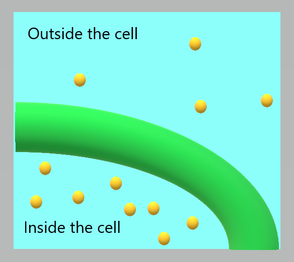
Passive transport occurs when substances cross the plasma membrane without any input of energy from the cell. No energy is required because the substances are moving from an area where they have a higher concentration to an area where they have a lower concentration. Concentration refers to the number of particles of a substance per unit of volume. The more particles of a substance in a given volume, the higher the concentration. A substance always moves from an area where it is more concentrated to an area where it is less concentrated.
There are several different types of passive transport, including simple diffusion, osmosis, and facilitated diffusion. Each type is described below.
SIMPLE DIFFUSION
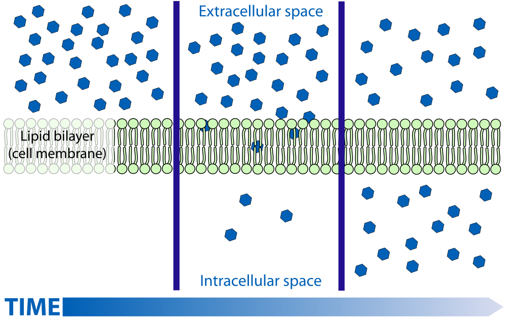
Diffusion is the movement of a substance due to a difference in concentration. It happens without any help from other molecules. The substance simply moves from the area where it is more concentrated to the area where it is less concentrated. Picture someone spraying perfume in the corner of a room. Do the perfume molecules stay in the corner? No, they spread out or diffuse throughout the room until they are evenly spread out. Figure 3.20 shows how diffusion works across a cell membrane Substances that can squeeze between the lipid molecules in the plasma membrane by simple diffusion are generally very small, hydrophobic molecules, such as molecules of oxygen and carbon dioxide.
Figure 3.20 Molecules diffuse across a membrane from an area of higher concentration to an area of lower concentration until the concentration is the same on both sides of the membrane.
Figure 3.21 Osmosis is a type of diffusion in which only water can cross the plasma membrane.
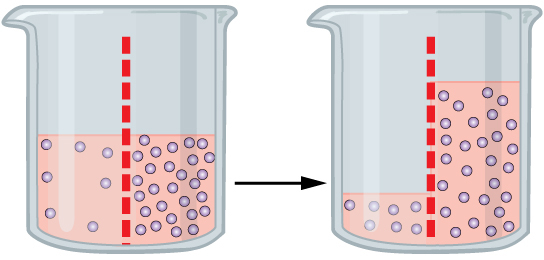
OSMOSIS
Osmosis is a special type of diffusion — the diffusion of water molecules across a membrane. Like other molecules, water moves from an area of higher concentration to an area of lower concentration. Water moves in or out of a cell until its concentration is the same on both sides of the plasma membrane. In Figure 3.21, the dotted red line shows a semi-permeable membrane. In the first beaker, there is an uneven concentration of solutes on either side of the membrane, but the solute cannot cross — diffusion of the solute can’t occur. In this case, water will move to even out the concentration as has happened on the beaker on the right side. The water levels are uneven, but the process of osmosis has evened out the concentration gradient.
FACILITATED DIFFUSION
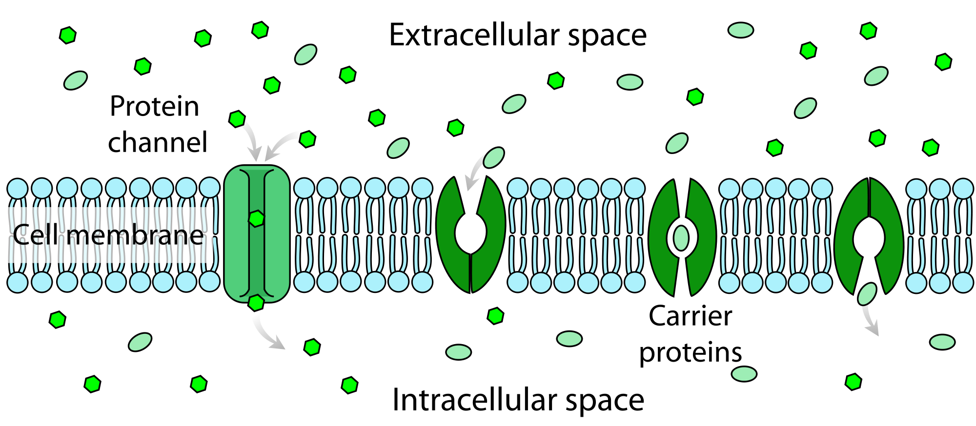
Water and many other substances cannot simply diffuse across a membrane. Hydrophilic molecules, charged ions, and relatively large molecules (such as glucose) all need help with diffusion. This help comes from special proteins in the membrane known as transport proteins. Diffusion with the help of transport proteins is called facilitated diffusion. There are several types of transport proteins, including channel proteins and carrier proteins. Both are shown in Figure 3.22.
Channel proteins form pores (or tiny holes) in the membrane. This allows water molecules and small ions to pass through the membrane without coming into contact with the hydrophobic tails of the lipid molecules in the interior of the membrane.
Carrier proteins bind with specific ions or molecules. In doing so, they change shape. As carrier proteins change shape, they carry the ions or molecules across the membrane.
Figure 3.22 Facilitated diffusion across a cell membrane. Channel proteins and carrier proteins help substances diffuse across a cell membrane. In this diagram, the channel and carrier proteins are helping substances move into the cell (from the extracellular space to the intracellular space).
TRANSPORT AND HOMEOSTASIS
For a cell to function normally, the inside of it must maintain a stable state. The concentrations of salts, nutrients, and other substances must be kept within certain ranges. The state in which stable conditions are maintained inside a cell (or an entire organism) is called homeostasis. Homeostasis requires constant adjustments, because conditions are always changing both inside and outside the cell. The transport of substances into and out of cells as described in this section plays an important role in homeostasis. By allowing the movement of substances into and out of cells, transport keeps conditions within normal ranges inside the cells and throughout the organism as a whole.
Watch this video “Cell Transport,” by the Amoeba Sisters:
Cell Transport with the Amoeba Sisters, 2016.
Review
What is the main difference between passive and active transport?
Summarize three different ways that passive transport can occur. Give an example of a substance that is transported in each way.
Explain how transport across the plasma membrane is related to homeostasis of the cell.
In general, why can only very small, hydrophobic molecules cross the cell membrane by simple diffusion?
Explain how facilitated diffusion assists with osmosis in cells. Define osmosis and facilitated diffusion in your answer.
Imagine a hypothetical cell with a higher concentration of glucose inside the cell than outside. Answer the following questions about this cell, assuming all transport across the membrane is passive, not active.
Can the glucose simply diffuse across the cell membrane? Why or why not?
Assuming that there are glucose transport proteins in the cell membrane, which way would glucose flow — into or out of the cell? Explain your answer.
If the concentration of glucose was equal inside and outside of the cell, do you think there would be a net flow of glucose across the cell membrane in one direction or the other? Explain your answer.
What are the similarities and differences between channel proteins and carrier proteins?
ACTIVE TRANSPORT
Figure 3.23 The Humvee challenge – Active transport.
LIKE PUSHING A HUMVEE UPHILL
You can tell by their faces that these airmen (Figure 3.23) are expending a lot of energy trying to push this Humvee up a slope. The men are participating in a competition that tests their brute strength against that of other teams. The Humvee weighs about 13 thousand pounds (about 5,897 kilograms), so it takes every ounce of energy they can muster to move it uphill against the force of gravity. Transport of some substances across a plasma membrane is a little like pushing a Humvee uphill — it can’t be done without adding energy.
WHAT IS ACTIVE TRANSPORT?
Some substances can pass into or out of a cell across the plasma membrane without any energy required because they are moving from an area of higher concentration to an area of lower concentration. This type of transport is called passive transport. Other substances require energy to cross a plasma membrane, often because they are moving from an area of lower concentration to an area of higher concentration, against the concentration gradient. This type of transport is called active transport. The energy for active transport comes from the energy-carrying molecule called ATP (adenosine triphosphate). Active transport may also require proteins called pumps, which are embedded in the plasma membrane. Two types of active transport are membrane pumps (such as the sodium-potassium pump) and vesicle transport.
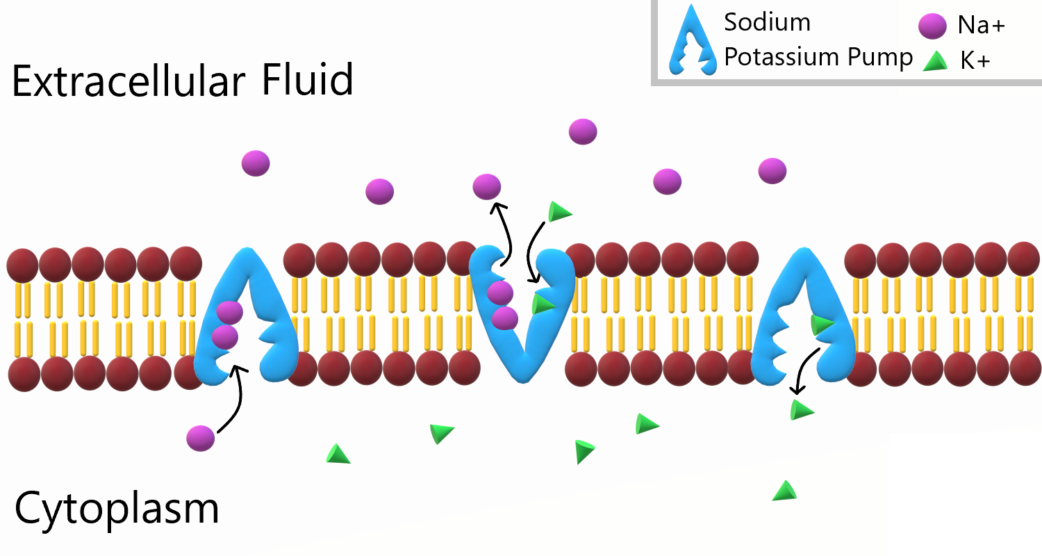
THE SODIUM-POTASSIUM PUMP
The sodium-potassium pump is a mechanism of active transport that moves sodium ions out of the cell and potassium ions into the cells — in all the trillions of cells in the body! Both ions are moved from areas of lower to higher concentration, so energy is needed for this “uphill” process. The energy is provided by ATP. The sodium-potassium pump also requires carrier proteins. Carrier proteins bind with specific ions or molecules, and in doing so, they change shape. As carrier proteins change shape, they carry the ions or molecules across the membrane. Figure 3.24 shows in greater detail how the sodium-potassium pump works, as well as the specific roles played by carrier proteins in this process.
Figure 3.24 The sodium-potassium pump moves sodium ions (Na+) out of the cell and potassium ions (K+) into the cell. First, three sodium ions bind with a carrier protein in the cell membrane. The carrier protein then changes shape, powered by energy from ATP, and as it does, it pumps the three sodium ions out of the cell. At that point, two potassium ions bind to the carrier protein. The process is reversed, and the potassium ions are pumped into the cell.
To appreciate the importance of the sodium-potassium pump, you need to know more about the roles of sodium and potassium in the body. Both are essential dietary minerals. You need to get them from the foods you eat. Both sodium and potassium are also electrolytes, which means they dissociate into ions (charged particles) in solution, allowing them to conduct electricity. Normal body functions require a very narrow range of concentrations of sodium and potassium ions in body fluids, both inside and outside of cells.
Sodium is the principal ion in the fluid outside of cells. Normal sodium concentrations are about ten times higher outside of cells than inside of cells. To move sodium out of the cell is moving it against the concentration gradient
Potassium is the principal ion in the fluid inside of cells. Normal potassium concentrations are about 30 times higher inside of cells than outside of cells. To move potassium into the cell is moving it against the concentration gradient.
Movement of these ions is one mechanism used by cells to control water volume.
These differences in concentration create an electrical and chemical gradient across the cell membrane called the membrane potential. Tightly controlling the membrane potential is critical for vital body functions, including the transmission of nerve impulses and contraction of muscles. A large percentage of the body’s energy goes to maintaining this potential across the membranes of its trillions of cells with the sodium-potassium pump.
VESICLE TRANSPORT
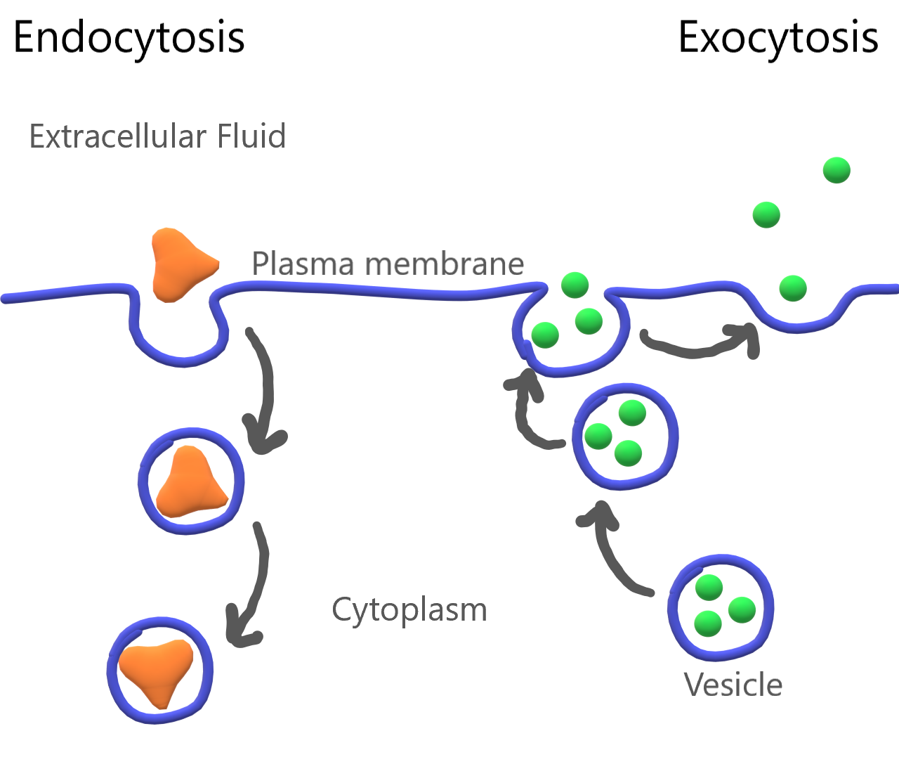
Some molecules, such as proteins, are too large to pass through the plasma membrane, regardless of their concentration inside and outside the cell. Very large molecules cross the plasma membrane with a different sort of help, called vesicle transport. Vesicle transport requires energy input from the cell, so it is also a form of active transport. There are two types of vesicle transport: endocytosis and exocytosis. Both types are shown in Figure 3.25.
Figure 3.25 Large molecules can enter and exit the cell with the help of vesicles. On the left side of the diagram you can see exocytosis, as large molecules exit the cell through the plasma membrane. On the right side of the diagram you can see endocytosis, as large molecules enter the cell through the plasma membrane, via vesicle formation.
Endocytosis
Endocytosis is a type of vesicle transport that moves a substance into the cell. The plasma membrane completely engulfs the substance, a vesicle pinches off from the membrane, and the vesicle carries the substance into the cell. When an entire cell or other solid particle is engulfed, the process is called phagocytosis. When fluid is engulfed, the process is called pinocytosis
Exocytosis
Exocytosis is a type of vesicle transport that moves a substance out of the cell (exo-, like “exit”). A vesicle containing the substance moves through the cytoplasm to the cell membrane. Because the vesicle membrane is a phospholipid bilayer. like the plasma membrane, the vesicle membrane fuses with the cell membrane, and the substance is released outside the cell.
Figure 3.26 Endocytosis brings substances into the cell via vesicle formation. Exocytosis allows substances to exit the cell by merging a transport vesicle with the cell membrane.
Feature: My Human Body
Maintaining the proper balance of sodium and potassium in body fluids by active transport is necessary for life itself, so it’s no surprise that getting the right balance of sodium and potassium in the diet is important for good health. Imbalances may increase the risk of high blood pressure, heart disease, diabetes, and other disorders.
If you are like the majority of North Americans, sodium and potassium are out of balance in your diet. You are likely to consume too much sodium and too little potassium. Follow these guidelines to help ensure that these minerals are balanced in the foods you eat:
Total sodium intake should be less than 2,300 mg/day. Most salt in the diet is found in processed foods, or added with a saltshaker. Stop adding salt and start checking food labels for sodium content. Foods considered low in sodium have less than 140 mg/serving (or 5 per cent daily value).
Total potassium intake should be 4,700 mg/day. It’s easy to add potassium to the diet by choosing the right foods — and there are plenty of choices! Most fruits and vegetables are high in potassium. Potatoes, bananas, oranges, apricots, plums, leafy greens, tomatoes, lima beans, and avocado are especially good sources. Other foods with substantial amounts of potassium are fish, meat, poultry, and whole grains. The collage below shows some of these potassium-rich foods.
Homeostasis and Cell Function
For a cell to function normally, a stable state must be maintained inside the cell. For example, the concentration of salts, nutrients, and other substances must be kept within a certain range. This helps a cell control water volume. If too much water enters the cell, it can burst. If there is too little water inside the cell, it will shrivel up and ide. The process of maintaining stable conditions inside a cell (or an entire organism) is homeostasis. Homeostasis requires constant adjustments because conditions are always changing both inside and outside the cell. By moving substances into and out of cells, they keep conditions within normal ranges inside the cells and the organism as a whole. There are two mechanisms by which cells maintain a stable state: the sodium-potassiumpump (explained above) and tonicity.
Tonicity
Tonicity describes how an extracellular solution can change the volume of a cell by affecting osmosis. A solution’s tonicity often directly correlates with the osmolarity of the solution. Osmolarity describes the total solute concentration of the solution. A solution with low osmolarity has a greater number of water molecules relative to the number of solute particles; a solution with high osmolarity has fewer water molecules with respect to solute particles. In a situation in which solutions of two different osmolarities are separated by a membrane permeable to water, though not to the solute, water will move from the side of the membrane with lower osmolarity (and more water) to the side with higher osmolarity (and less water). This effect makes sense if you remember that the solute cannot move across the membrane, and thus the only component in the system that can move—the water—moves along its own concentration gradient. An important distinction that concerns living systems is that osmolarity measures the number of particles (which may be molecules) in a solution. Therefore, a solution that is cloudy with cells may have a lower osmolarity than a solution that is clear if the second solution contains more dissolved molecules than there are cells.
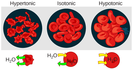
Hypotonic Solutions
Three terms—hypotonic, isotonic, and hypertonic—are used to relate the osmolarity of a cell to the osmolarity of the extracellular fluid that contains the cells. In a hypotonic situation, the extracellular fluid has lower osmolarity than the fluid inside the cell, and water enters the cell. (In living systems, the point of reference is always the cytoplasm, so the prefix hypo- means that the extracellular fluid has a lower concentration of solutes, or a lower osmolarity, than the cell cytoplasm. ) It also means that the extracellular fluid has a higher concentration of water in the solution than does the cell. In this situation, water will follow its concentration gradient and enter the cell, causing the cell to expand.
Figure 3.27: Changes in Cell Shape Due to Dissolved Solutes: Osmotic pressure changes the shape of red blood cells in hypertonic, isotonic, and hypotonic solutions.
Hypertonic Solutions
As for a hypertonic solution, the prefix hyper- refers to the extracellular fluid having a higher osmolarity than the cell’s cytoplasm; therefore, the fluid contains less water than the cell does. Because the cell has a relatively higher concentration of water, water will leave the cell, and the cell will shrink.
Isotonic Solutions
In an isotonic solution, the extracellular fluid has the same osmolarity as the cell. If the osmolarity of the cell matches that of the extracellular fluid, there will be no net movement of water into or out of the cell, although water will still move in and out.
Blood cells and plant cells in hypertonic, isotonic, and hypotonic solutions take on characteristic appearances. Cells in an isotonic solution retain their shape. Cells in a hypotonic solution swell as water enters the cell and may burst if the concentration gradient is large enough between the inside and outside of the cell. Cells in a hypertonic solution shrink as water exits the cell, becoming shriveled.
Review
Define active transport.
What is the sodium-potassium pump? Why is it so important?
The drawing below shows the fluid inside and outside of a cell. The dots represent molecules of a substance needed by the cell. Explain which type of transport — active or passive — is needed to move the molecules into the cell.
What are the similarities and differences between phagocytosis and pinocytosis?
What is the functional significance of the shape change of the carrier protein in the sodium-potassium pump after the sodium ions bind?
A potentially deadly poison derived from plants called ouabain blocks the sodium-potassium pump and prevents it from working. What do you think this does to the sodium and potassium balance in cells? Explain your answer.
What two mechanisms control a cell’s volume?
Distinguish between isotonic, hypertonic and hypotonic solutions.
In the scientific world, energy is defined as the ability to do work. You can often see energy at work in living things — a bird flies through the air, a firefly glows in the dark, a dog wags its tail. These are obvious ways that living things use energy, but living things constantly use energy in less obvious ways, as well. The chemical energy that organisms need comes from food. Food consists of organic molecules, such as triglycerides and glycogen, that store energy in their chemical bonds.
Inside every cell of all living things, energy is needed to carry out life processes. Metabolism is the set of life-sustaining chemical processes that enables organisms transform the chemical energy stored in molecules into energy that can be used for cellular processes. Energy is required to break down (Catabolism) and build up (Anabolism) molecules, and to transport many molecules across plasma membranes. All of life’s work needs energy. A lot of energy is also simply lost to the environment as heat. The story of life is a story of energy flow — its capture, its change of form, its use for work, and its loss as heat. Energy (unlike matter) cannot be recycled, so organisms require a constant input of energy. Life runs on chemical energy. Where do living organisms get this chemical energy?
Metabolic Pathways
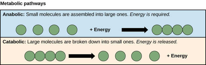
The processes of making and breaking down carbohydrate molecules illustrate two types of metabolic pathways. A metabolic pathway is a step-by-step series of interconnected biochemical reactions that convert a substrate molecule or molecules through a series of metabolic intermediates, eventually yielding a final product or products. For example, one metabolic pathway for carbohydrates breaks large molecules down into glucose. Another metabolic pathway might build glucose into large carbohydrate molecules for storage. The first of these processes requires energy and is referred to as anabolic. The second process produces energy and is referred to as catabolic. Consequently, metabolism is composed of these two opposite pathways:
Anabolism (building molecules)
Catabolism (breaking down molecules)
Figure 3.28: Anabolic and catabolic pathways: Anabolic pathways are those that require energy to synthesize larger molecules. Catabolic pathways are those that generate energy by breaking down larger molecules. Both types of pathways are required for maintaining the cell’s energy balance.
Anabolic Pathways
Anabolic pathways require an input of energy to synthesize complex molecules from simpler ones. One example of an anabolic pathway is the synthesis of sugar from CO2. Other examples include the synthesis of large proteins from amino acid building blocks and the synthesis of new DNA strands from nucleic acid building blocks. These processes are critical to the life of the cell, take place constantly, and demand energy provided by ATP and other high-energy molecules like NADH (nicotinamide adenine dinucleotide) and NADPH.
Catabolic Pathways
Catabolic pathways involve the degradation of complex molecules into simpler ones, releasing the chemical energy stored in the bonds of those molecules. Some catabolic pathways can capture that energy to produce ATP, the molecule used to power all cellular processes. Other energy-storing molecules, such as lipids, are also broken down through similar catabolic reactions to release energy and make ATP.
Importance of Enzymes
Chemical reactions in metabolic pathways rarely take place spontaneously. Each reaction step is facilitated, or catalyzed, by a protein called an enzyme. Enzymes are important for catalyzing all types of biological reactions: those that require energy as well as those that release energy.
Energy
Organisms mainly use two types of molecules for chemical energy: glucose and ATP. Both molecules are used as fuels throughout the living world.
Glucose is a simple carbohydrate with the chemical formula C6H12O6. It stores chemical energy in a concentrated, stable form. In your body, glucose is the form of energy that is carried in your blood and taken up by each of your trillions of cells. Glucose is the end product of photosynthesis and it is the nearly universal food for life. In Figure 3.29, you can see how photosynthesis stores energy from the sun in the glucose molecule and then how cellular respiration breaks the bonds in glucose to retrieve the energy.
Figure 3.29 Energy transfer in photosynthesis and cellular respiration.
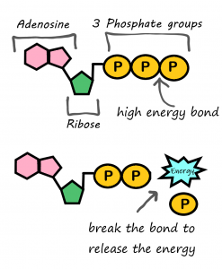
ATP (adenosine triphosphate) is the energy-carrying molecule that cells use to power most cellular processes (nerve impulse conduction, protein synthesis and active transport are good examples of cell processes that rely on ATP as their energy source). ATP is made during the first half of photosynthesis and then used for energy during the second half of photosynthesis, when glucose is made. ATP releases energy when it gives up one of its three phosphate groups (Pi) and changes to ADP (adenosine diphosphate, which has two phosphate groups), as shown in Figure 3.30. Thus, the breakdown of ATP into ADP + Pi is a catabolic reaction that releases energy (exothermic). ATP is made from the combination of ADP and Pi, an anabolic reaction that takes in energy (endothermic).
WHY ORGANISMS NEED BOTH GLUCOSE AND ATP
Why do living things need glucose if ATP is the molecule that cells use for energy? A molecule of glucose contains more chemical energy in a smaller “package” than a molecule of ATP. Glucose is also more stable than ATP. Therefore, glucose is better for storing and transporting energy. Glucose, however, is too powerful for cells to use. ATP, on the other hand, contains just the right amount of energy to power life processes within cells. For these reasons, both glucose and ATP are needed by living things.
Figure 3.30 ATP (adenosine TRI phosphate) can be converted to ADP (adenosine DI phosphate) to release the energy stored in the chemical bonds between the second and third phosphate group.
3.9 CELLULAR RESPIRATION
The flow of energy through living organisms begins with photosynthesis. This process stores energy from sunlight in the chemical bonds of glucose. By breaking the chemical bonds in glucose, cells release the stored energy and make the ATP they need. The process in which glucose is broken down and ATP is made is called cellular respiration.
WHAT IS CELLULAR RESPIRATION?
Cellular respiration is the process by which living cells break down glucose molecules and release energy. The process is similar to burning, although it doesn’t produce light or intense heat as a campfire does. This is because cellular respiration releases the energy in glucose slowly andin many small steps. It uses the energy released to form molecules of ATP, the energy-carrying molecules that cells use to power biochemical processes. In this way, cellular respiration is an example of energy coupling: glucose is broken down in an exothermic reaction, and then the energy from this reaction powers the endothermic reaction of the formation of ATP. Cellular respiration involves many chemical reactions, but they can all be summed up with this chemical equation:
C6H12O6 6O2 → 6CO2 6H2O Chemical Energy (in ATP)
In words, the equation shows that glucose (C6H12O6) and oxygen (O2) react to form carbon dioxide (CO2) and water (H2O), releasing energy in the process. Because oxygen is required for cellular respiration, it is an aerobic process.
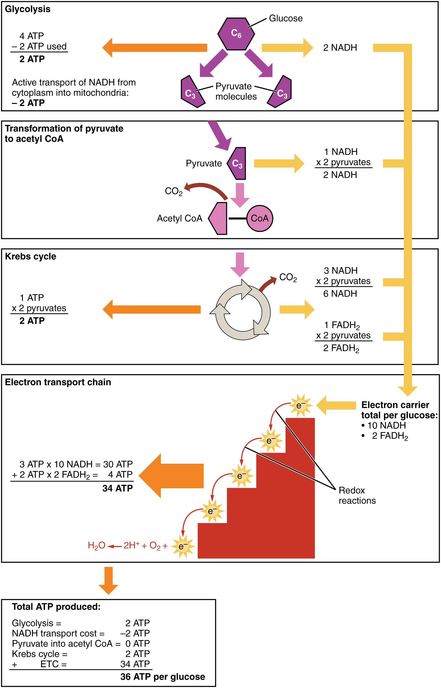
Cellular respiration occurs in the cells of all living things. All of them burn glucose to form ATP. The reactions of cellular respiration can be grouped into three stages: glycolysis, the Krebs cycle (also called the citric acid cycle), and electron transport. Figure 3.31 gives an overview of these three stages, which are also described in detail below.
Figure 3.31 Cellular respiration takes place in the stages shown here. The process begins with a molecule of glucose, which has six carbon atoms. What happens to each of these atoms of carbon?
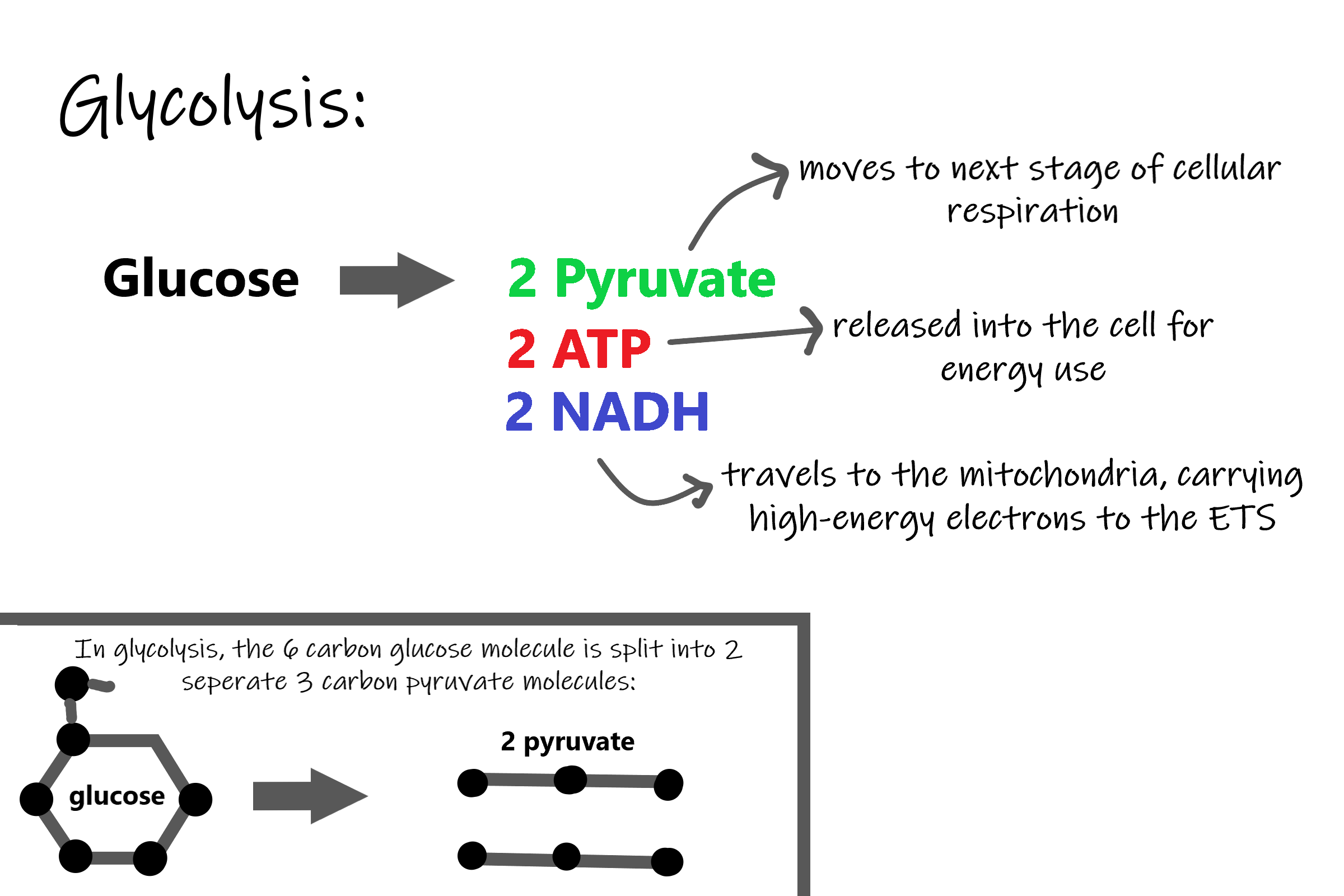
CELLULAR RESPIRATION STAGE I: GLYCOLYSIS
The first stage of cellular respiration is glycolysis which happens in the cytosol of the cytoplasm.
SPLITTING GLUCOSE
The word glycolysis literally means “glucose splitting,” which is exactly what happens in this stage. Enzymes split a molecule of glucose into two molecules of pyruvate (also known as pyruvic acid). This occurs in several steps, as summarized in the following diagram.
Figure 3.32 Glycolysis is a complex ten-step reaction that ultimately converts glucose into two molecules of pyruvate. This releases energy, which is transferred to ATP. How many ATP molecules are made during this stage of cellular respiration?
RESULTS OF GLYCOLYSIS
Energy is needed at the start of glycolysis to split the glucose molecule into two pyruvate molecules which go on to stage II of cellular respiration. The energy needed to split glucose is provided by two molecules of ATP; this is called the energy investment phase. As glycolysis proceeds, energy is released, and the energy is used to make four molecules of ATP; this is the energy harvesting phase. As a result, there is a net gain of two ATP molecules during glycolysis. During this stage, high-energy electrons are also transferred to molecules of NAD to produce two molecules of NADH, another energy-carrying molecule. NADH is used in stage III of cellular respiration to make more ATP.
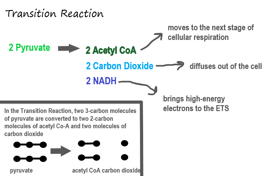
TRANSITION REACTION (PREPARATORY STEP)
Before pyruvate can enter the next stage of cellular respiration it needs to be modified slightly. The transition reaction is a very short reaction which converts the two molecules of pyruvate to two molecules of acetyl CoA, carbon dioxide, and two high energy electron pairs convert NAD to NADH. The carbon dioxide is released, the acetyl CoA moves to the mitochondria to enter the Kreb’s Cycle (stage II), and the NADH carries the high energy electrons to the Electron Transport System (stage III).
Figure 3.33: During the Transition Reaction, pyruvate is converted to acetyl CoA and carbon dioxide.
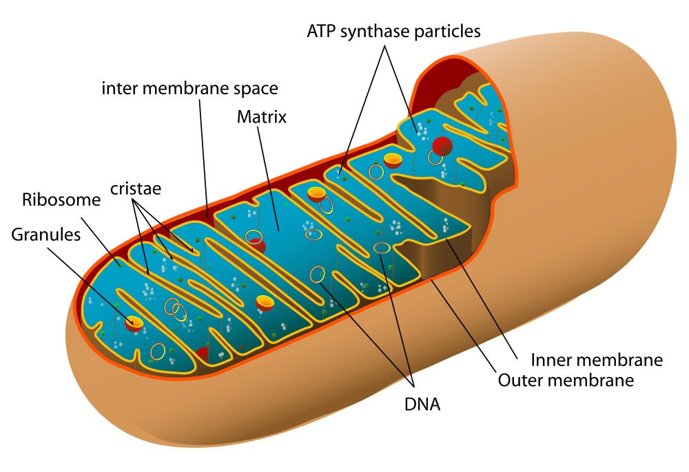
STRUCTURE OF THE MITOCHONDRION
Before you read about the last two stages of cellular respiration, you need to know more about the mitochondrion, where these two stages take place. A diagram of a mitochondrion is shown in Figure 3.34.
The structure of a mitochondrion is defined by an inner and outer membrane. This structure plays an important role in aerobic respiration.
As you can see from the figure, a mitochondrion has an inner and outer membrane. The space between the inner and outer membrane is called the intermembrane space. The space enclosed by the inner membrane is called the matrix. The second stage of cellular respiration (the Krebs cycle) takes place in the matrix. The third stage (electron transport) happens on the inner membrane.
Figure 3.34 Labelled mitochondrion structure.
CELLULAR RESPIRATION STAGE II: THE KREBS CYCLE
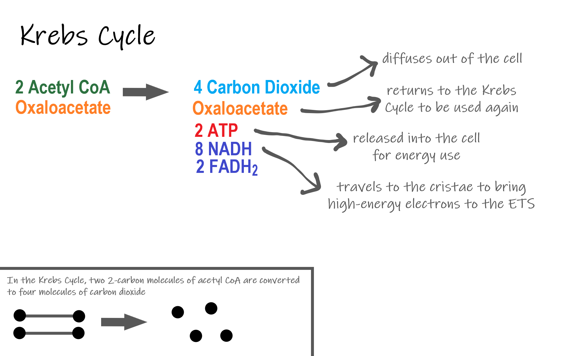
Recall that glycolysis produces two molecules of pyruvate (pyruvic acid), which are then converted to acetyl CoA during the short transition reaction. These molecules enter the matrix of a mitochondrion, where they start the Krebs Cycle (also known as the Citric Acid Cycle). The reason this stage is considered a cycle is because a molecule called oxaloacetate is present at both the beginning and end of this reaction and is used to break down the two molecules of acetyl CoA. The reactions that occur next are shown in Figure 3.35.
Figure 3.35Reactants and products of the Krebs Cycle.
Steps of the Krebs Cycle
The Krebs cycle itself actually begins when acetyl-CoA combines with a four-carbon molecule called OAA (oxaloacetate) (see Figure 3.35). This produces citric acid, which has six carbon atoms. This is why the Krebs cycle is also called the citric acid cycle.
After citric acid forms, it goes through a series of reactions that release energy. The energy is captured in molecules of NADH, ATP, and FADH2, another energy-carrying coenzyme. Carbon dioxide is also released as a waste product of these reactions.
The final step of the Krebs cycle regenerates OAA, the molecule that began the Krebs cycle. This molecule is needed for the next turn through the cycle. Two turns are needed because glycolysis produces two pyruvic acid molecules when it splits glucose.
RESULTS OF THE GLYCOLYSIS, TRANSITION REACTION AND KREBS CYCLE
After glycolysis, transition reaction, and the Krebs cycle, the glucose molecule has been broken down completely. All six of its carbon atoms have combined with oxygen to form carbon dioxide. The energy from its chemical bonds has been stored in a total of 16 energy-carrier molecules. These molecules are:
4 ATP (2 from glycolysis, 2 from Krebs Cycle)
12 NADH (2 from glycolysis, 2 from transition reaction, and 8 from Krebs cycle)
2 FADH2 (both from the Krebs cycle)
The events of cellular respiration up to this point are exergonic reactions – they are releasing energy that had been stored in the bonds of the glucose molecule. This energy will be transferred to the third and final stage of cellular respiration: the Electron Transport System, which is an endergonic reactions.
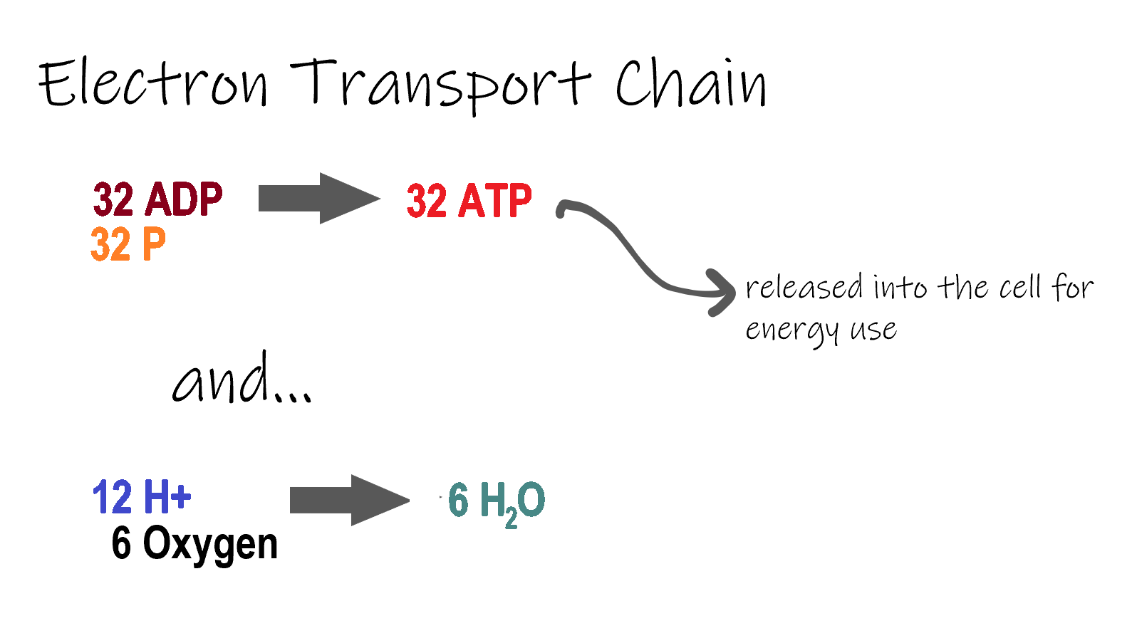
CELLULAR RESPIRATION STAGE III: ELECTRON TRANSPORT CHAIN
Figure 3.36. Reactants and products of the electron transport chain.
ETC, the final stage in cellular respiration produces 32 ATP. The Electron Transport Chain is the final stage of cellular respiration. In this stage, energy being transported by NADH and FADH2 is transferred to ATP. In addition, oxygen acts as the final proton acceptor for the hydrogens released from all the NADH and FADH2, forming water. Figure 3.36 shows the reactants and products of the ETC.
TRANSPORTING ELECTRONS
The Electron transport chain is the third stage of cellular respiration and is illustrated in Figure 3.37. During this stage, high-energy electrons are released from NADH and FADH2, and they move along electron-transport chains on the inner membrane of the mitochondrion. An electron-transport chain is a series of molecules that transfer electrons from molecule to molecule by chemical reactions. Some of the energy from the electrons is used to pump hydrogen ions (H ) across the inner membrane, from the matrix into the intermembrane space. This ion transfer creates an electrochemical gradient that drives the synthesis of ATP.
Figure 3.37 Electron-transport chains on the inner membrane of the mitochondrion carry out the last stage of cellular respiration.
Making ATP
As shown in Figure 3.37, the pumping of hydrogen ions across the inner membrane creates a greater concentration of the ions in the intermembrane space than in the matrix. This gradient causes the ions to flow back across the membrane into the matrix, where their concentration is lower. ATP synthase acts as a channel protein, helping the hydrogen ions cross the membrane. It also acts as an enzyme, forming ATP from ADP and inorganic phosphate in a process called oxidative phosphorylation. After passing through the electron-transport chain, the “spent” electrons combine with oxygen to form water.
HOW MUCH ATP?
You have seen how the three stages of aerobic respiration use the energy in glucose to makeATP. How much ATP is produced in all three stages combined? Glycolysis produces two ATP molecules, and the Krebs cycle produces two more. Electron transport begins with several molecules of NADH and FADH2 from the Krebs cycle and transfers their energy into as many as 34 more ATP molecules. All told, then, up to 38 molecules of ATP can be produced from just one molecule of glucose in the process of cellular respiration.
Review
What is the purpose of cellular respiration? Provide a concise summary of the process.
State what happens during glycolysis.
Describe the structure of a mitochondrion.
What molecule is present at both the beginning and end of the Krebs cycle?
What happens during the electron transport stage of cellular respiration?
How many molecules of ATP can be produced from one molecule of glucose during all three stages of cellular respiration combined?
Do plants undergo cellular respiration? Why or why not?
Explain why the process of cellular respiration described in this section is considered aerobic.
Name three energy-carrying molecules involved in cellular respiration.
Which stage of aerobic cellular respiration produces the most ATP?
Cellular Respiration and the Mighty Mitochondria, The Amoeba Sisters, 2014.
3.10 ANAEROBIC PROCESSES
Figure 3.38 Sprinters racing on a track.
FAST AND FURIOUS
These sprinters’ muscles will need a lot of energy to complete this short race, because they will be running at top speed. The action won’t last long, but it will be very intense. The energy each sprinter needs can’t be provided quickly enough by aerobic cellular respiration. Instead, their muscle cells must use a different process to power their activity.
MAKING ATP WITHOUT OXYGEN
Living things’ cells power their activities with the energy-carrying molecule ATP (adenosine triphosphate). The cells of most living things make ATP from glucose in the process of cellular respiration. This process occurs in three stages: glycolysis the Krebs cycle and electron transport. The latter two stages require oxygen, making cellular respiration anaerobic process. When oxygen is not available in cells, the ETS quickly shuts down. Luckily, there are also ways of making ATP from glucose which are anaerobic, which means that they do not require oxygen. These processes are referred to collectively as anaerobic respiration.
FERMENTATION
One important way of making ATP without oxygen is fermentation. Fermentation starts with glycolysis, which does not require oxygen, but it does not involve the latter two stages of aerobic cellular respiration (the Krebs cycle and electron transport. There are two types of fermentation: alcoholic fermentation and lactic acid fermentation. We make use of both types of fermentation using other organisms, but only lactic acid fermentation actually takes place inside the human body.
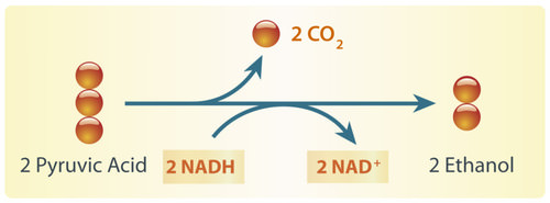
ALCOHOLIC FERMENTATION
Figure 3.39 In alcoholic fermentation, pyruvate is converted to ethanol and carbon dioxide. During this process, NAD+ is formed, which allows glycolysis to continue making ATP.
Alcoholic fermentation is carried out by single-celled fungi (called yeasts), as well as some bacteria. We use alcoholic fermentation in these organisms to make biofuels, bread, and wine. The biofuel ethanol (a type of alcohol), for example, is produced by alcoholic fermentation of the glucose in corn or other plants. The process by which this happens is summarized in the diagram below. The two pyruvic acid molecules shown in the diagram come from the splitting of glucose in the first stage of the process (glycolysis). ATP is also made during glycolysis. Two molecules of ATP are produced from each molecule of glucose.
Figure 3.40 Holes in bread created by carbon dioxide.
Yeasts in bread dough also use alcoholic fermentation for energy. They produce carbon dioxide gas as a waste product. The carbon dioxide released causes bubbles in the dough and explains why the dough rises. Do you see the small holes in the bread pictured to the right? The holes were formed by bubbles of carbon dioxide gas.
As you have probably guessed, yeast is also used in producing alcoholic beverages. When making beer, brewers will add yeast to a mix of barley and hops. In the absence of oxygen, yeast will carry out alcoholic fermentation in order to convert the glucose in the barley into energy, producing the alcohol content as well as the carbonation present in beer.
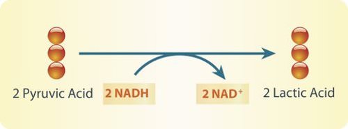
Lactic Acid Fermentation
Lactic Acid fermentation is carried out by certain bacteria, including the bacteria in yogurt. It is also carried out by your muscle cells when you work them hard and fast. This is how the muscles of the sprinters pictured above get energy for their short-lived — but intense — activity. When this happens, your muscles are using ATP faster than your cardiovascular system can deliver oxygen! The process by which this happens is summarized in the diagram below. Again, the two pyruvic acid molecules shown in the diagram come from the splitting of glucose in the first stage of the process (glycolysis). It is also during this stage that two ATP molecules are produced. The rest of the processes produce lactic acid. Note that, unlike in alcoholic fermentation, there is no carbon dioxide waste product in lactic acid fermentation.
Figure 3.41 Lactic acid fermentation formula.
Lactic acid fermentation produces lactic acid and NAD+. The NAD+ cycles back to allow glycolysis to continue so more ATP is made. Each circle represents a carbon atom.
Did you ever run a race, lift heavy weights, or participate in some other intense activity and notice that your muscles start to feel a burning sensation? This may occur when your muscle cells use lactic acid fermentation to provide ATP for energy. The buildup of lactic acid in the muscles causes a burning feeling. This painful sensation is useful if it gets you to stop overworking your muscles and allow them a recovery period, during which cells can eliminate the lactic acid.
PROS AND CONS OF ANAEROBIC RESPIRATION
With oxygen, organisms can use aerobic cellular respiration to produce up to 38 molecules of ATP from just one molecule of glucose. Without oxygen, organisms must use anaerobic respiration to produce ATP, and this process produces only two molecules of ATP per molecule of glucose. Although anaerobic respiration produces less ATP, it has the advantage of doing so very quickly. For example, it allows your muscles to get the energy they need for short bursts of intense activity. Aerobic cellular respiration, in contrast, produces ATP more slowly.
FERMENTATION IN FOOD PRODUCTION
Anaerobic respiration is also used in the food industry. You read about yeast’s role in making bread and beer, but did you know that there are many microbes that are used to create the food we eat, including cheese, sour cream, yogurt, soy sauce, olives, pepperoni, and many more. Watch the video below to learn more about fermentation in the food industry.
The beneficial bacteria that make delicious food – Erez Garty, TED-Ed, 2016.
Review
Explain the primary difference between aerobic cellular respiration and anaerobic respiration.
What is fermentation?
Compare and contrast alcoholic and lactic acid fermentation.
Identify the major pros and the major cons of anaerobic respiration relative to aerobic cellular respiration.
What process is shared between aerobic cellular respiration and anaerobic respiration? Describe the process briefly. Why can this process happen in anaerobic respiration, as well as aerobic respiration?
CASE STUDY: MORE THAN JUST TIRED
We all get tired sometimes, especially if we have been doing a lot of physical activity. But for Jasmin (Figure 3.41), a 34-year-old former high school track star who is now a recreational runner, her tiredness was going far beyond what she thought should be normal for someone in generally good physical shape. She was experiencing extreme fatigue after her runs, as well as muscle cramping, spasms, and an unusual sense of heaviness in her legs. At first, she just chalked it up to getting older, but her exhaustion and pain worsened to the point where the former athlete could no longer run for more than a few minutes at a time. She began to experience other unusual symptoms, such as blurry vision and vomiting for no apparent reason.
Figure 3.42 Jasmin was feeling a level of fatigue that was far beyond normal tiredness.
Jasmin discovered that her extreme fatigue, muscle pain, vision problems, and vomiting were due to problems in her mitochondria. Mitochondria are small, membrane-bound organelles found in eukaryotic cells that provide energy for the cells of the body. They do this by carrying out the final two steps of aerobic cellular respiration: the Krebs cycle and electron transport. This is the major way that the human body breaks down the sugar glucose from food into a form of energy cells can use, namely the molecule ATP
Because mitochondria provide energy for cells, you can understand why Jasmin was experiencing extreme fatigue, particularly after running. Her damaged mitochondria could not keep up with her need for energy, particularly after intense exercise, which requires a lot of additional energy. What is perhaps not so obvious are the reasons for her other symptoms, such as blurry vision, muscle spasms, and vomiting. All of the cells in the body require energy in order to function properly. Mitochondrial diseases can cause problems in mitochondria in any cell of the body, including muscle cells and cells of the nervous system, which includes the brain and nerves. The nervous system and muscles work together to control vision and digestive system functions, such as vomiting, so when they are not functioning properly, a variety of symptoms can emerge. This also explains why Jasmin’s niece, who has a similar mitochondrial disease, has symptoms related to brain function, such as seizures and learning disabilities. Our cells are microscopic, and mitochondria are even tinier — but they are essential for the proper functioning of our bodies. When they are damaged, serious health effects can occur.
Figure 3.43 Mitochondrial disease can manifest itself very differently in different people, even if they are related. Jasmin and her niece have the same mitochondrial disease, but with different age of onset, different symptoms and different severity of symptoms.
One seemingly confusing aspect of mitochondrial diseases is that the type of symptoms, severity of symptoms, and age of onset can vary wildly between people — even within the same family! In Jasmin’s case, she did not notice symptoms until adulthood, while her niece had more severe symptoms starting at a much younger age. This makes sense when you know more about how mitochondrial diseases work.
Inherited mitochondrial diseases can be due to damage in either the DNA in the nucleus of cells or in the DNA in the mitochondria themselves. When the mitochondrial DNA is damaged (or mutated) it can result in some types of mitochondrial diseases. However, these mutations do not typically affect all of the mitochondria in a cell. During cell division, organelles such as mitochondria are replicated and passed down to the new daughter cells. If some of the mitochondria are damaged, and others are not, the daughter cells can have different amounts of damaged mitochondria. This helps explain the wide range of symptoms in people with mitochondrial diseases — even ones in the same family — because different cells in their bodies are affected in varying degrees. Jasmin’s niece was affected strongly, and her symptoms were noticed early, while Jasmin’s symptoms were milder and did not become apparent until adulthood.
There is still much more that needs to be discovered about the different types of mitochondrial diseases. But by learning about cells, their organelles, how they obtain energy, and how they divide, you should now have a better understanding of the biology behind these diseases.
Apply your understanding of cells to your own life. Can you think of other diseases that affect cellular structures or functions. Do they affect people you know? Since your entire body is made of cells, when cells are damaged or not functioning properly, it can cause a wide variety of health problems.
Attributions
This chapter is composed of text taken from of the following sources:
Gyurkovics, G. (2024). Human biology. CK-12 License. Authored, remixed, and/or curated by S. Wakim & M. Grewal. Edited to the style and standards of the LibreTexts platform. Retrieved from [Human Biology (Gabor Gyurkovics) – Biology LibreTexts]