WE ALL SCREAM FOR ICE CREAM
If you’re an ice cream lover, then just the sight of this yummy ice cream cone may make your mouth water. The “water” in your mouth is actually saliva, a fluid released by glands that are part of the digestive system. Saliva contains digestive enzymes, among other substances important for digestion. When your mouth waters at the sight of a tasty treat, it’s a sign that your digestive system is preparing to digest food.
WHAT IS THE DIGESTIVE SYSTEM?
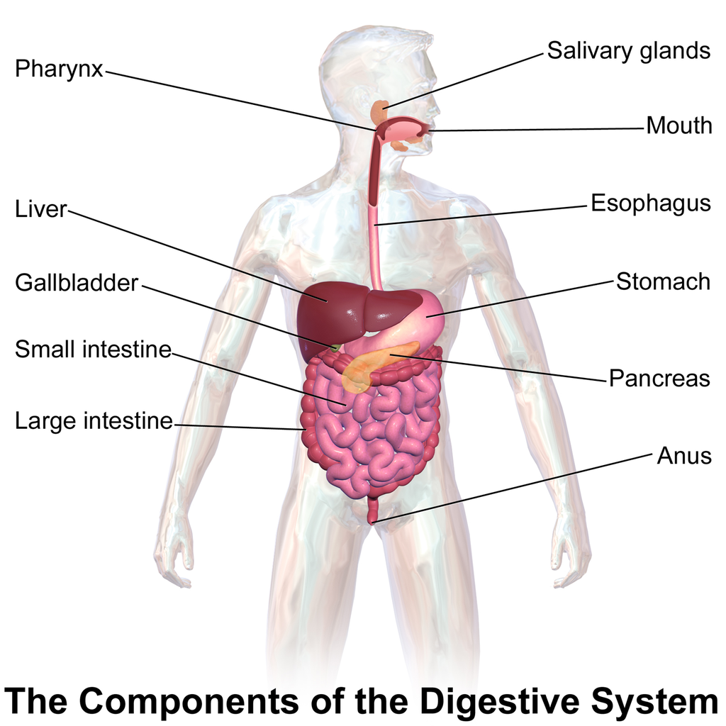
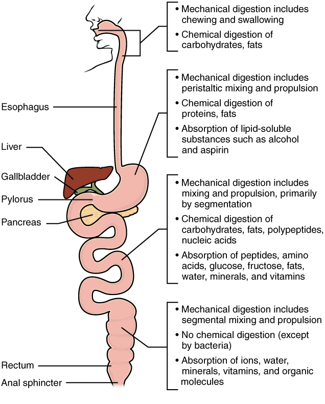
The digestive system consists of organs that break down food, absorb its nutrients, and expel any remaining waste. Organs of the digestive system are shown in Figure 13.2. Most of these organs make up the gastrointestinal (GI) tract, through which food actually passes. The rest of the organs of the digestive system are called accessory organs. These organs secrete enzymes and other substances into the GI tract, but food does not actually pass through them.
Functions of the Digestive System
The digestive system has three main functions relating to food: digestion of food, absorption of nutrients from food, and elimination of solid food waste. Digestion is the process of breaking down food into components the body can absorb. It consists of two types of processes: mechanical digestion and chemical digestion. Mechanical digestion is the physical breakdown of chunks of food into smaller pieces, and it takes place mainly in the mouth and stomach. Chemical digestion is the chemical breakdown of large, complex food molecules into smaller, simpler nutrient molecules that can be absorbed by body fluids (blood or lymph). This type of digestion begins in the mouth and continues in the stomach but occurs mainly in the small intestine.
After food is digested, the resulting nutrients are absorbed. Absorption is the process in which substances pass into the bloodstream or lymph system to circulate throughout the body. Absorption of nutrients occurs mainly in the small intestine. Any remaining matter from food that is not digested and absorbed passes out of the body through the anus in the process of elimination.
GASTROINTESTINAL TRACT
The gastrointestinal (GI) tract is basically a long, continuous tube that connects the mouth with the anus. If it were fully extended, it would be about nine meters long in adults. It includes the mouth, pharynx, esophagus, stomach, and small and large intestines. Food enters the mouth, and then passes through the other organs of the GI tract, where it is digested and/or absorbed. Finally, any remaining food waste leaves the body through the anus at the end of the large intestine. It takes up to 50 hours for food or food waste to make the complete trip through the GI tract.
TISSUES OF THE GI TRACT
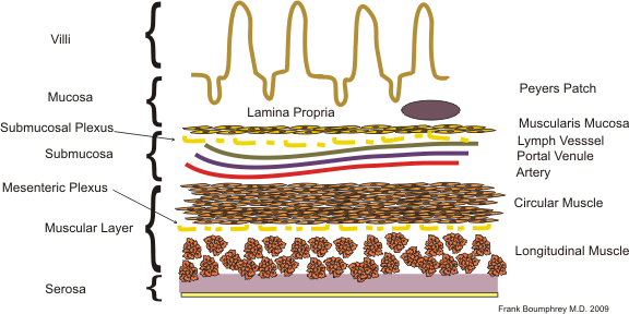
The walls of the organs of the GI tract consist of four different tissue layers, which are illustrated in Figure 13.3: mucosa, submucosa, muscularis externa, and serosa.
- The mucosa is the innermost layer surrounding the lumen (open space within the organs of the GI tract). This layer consists mainly of epithelium with the capacity to secrete and absorb substances. The epithelium can secret digestive enzymes and mucus, and it can absorb nutrients and water.
- The submucosa layer consists of connective tissue that contains blood and lymph vessels, as well as nerves. The vessels are needed to absorb and carry away nutrients after food is digested, and nerves help control the muscles of the GI tract organs.
- The muscularis externa layer contains two types of smooth muscle: longitudinal muscle and circular muscle. Longitudinal muscle runs the length of the GI tract organs, and circular muscle encircles the organs. Both types of muscles contract to keep food moving through the tract by the process of peristalsis, which is described below.
- The serosa layer is the outermost layer of the walls of GI tract organs. This is a thin layer that consists of connective tissue and separates the organs from surrounding cavities and tissues.
 |
 |
PERISTALISIS IN THE GI TRACT
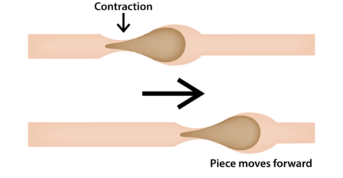
The muscles in the walls of GI tract organs enable peristalsis, which is illustrated in Figure 13.5. Peristalsis is a continuous sequence of involuntary muscle contraction and relaxation that moves rapidly along an organ like a wave, similar to the way a wave moves through a spring toy. Peristalsis in organs of the GI tract propels food through the tract.
Watch the video “What is peristalsis?” by Mister Science to see peristalsis in action:
What is peristalsis? Mister Science, 2018.
IMMUNE FUNCTION OF THE GI TRACT
The GI tract plays an important role in protecting the body from pathogens. The surface area of the GI tract is estimated to be about 32 square meters (105 square feet), or about half the area of a badminton court. This is more than three times the area of the exposed skin of the body, and it provides a lot of area for pathogens to invade the tissues of the body. The innermost mucosal layer of the walls of the GI tract provides a barrier to pathogens so they are less likely to enter the blood or lymph circulations. The mucus produced by the mucosal layer, for example, contains antibodies that mark many pathogenic microorganisms for destruction. Enzymes in some of the secretions of the GI tract also destroy pathogens. In addition, stomach acids have a very low pH that is fatal for many microorganisms that enter the stomach.
DIVISIONS OF THE GI TRACT
The GI tract is often divided into an upper GI tract and a lower GI tract. For medical purposes, the upper GI tract is typically considered to include all the organs from the mouth through the first part of the small intestine, called the duodenum. For our instructional purposes, it makes more sense to include the mouth through the stomach in the upper GI tract, and all of the small intestine — as well as the large intestine — in the lower GI tract.
Upper GI Tract
The mouth is the first digestive organ that food enters. The sight, smell, or taste of food stimulates the release of digestive enzymes and other secretions by salivary glands inside the mouth. The major salivary gland enzyme is amylase. It begins the chemical digestion of carbohydrates by breaking down starches into sugar. The mouth also begins the mechanical digestion of food. When you chew, your teeth break, crush, and grind food into increasingly smaller pieces. Your tongue helps mix the food with saliva and also helps you swallow.
A lump of swallowed food is called a bolus. The bolus passes from the mouth into the pharynx, and from the pharynx into the esophagus. The esophagus is a long, narrow tube that carries food from the pharynx to the stomach. It has no other digestive functions. Peristalsis starts at the top of the esophagus when food is swallowed and continues down the esophagus in a single wave, pushing the bolus of food ahead of it.
From the esophagus, food passes into the stomach, where both mechanical and chemical digestion continue. The muscular walls of the stomach churn and mix the food, thus completing mechanical digestion, as well as mixing the food with digestive fluids secreted by the stomach. One of these fluids is hydrochloric acid (HCl). In addition to killing pathogens in food, it gives the stomach the low pH needed by digestive enzymes that work in the stomach. One of these enzymes is pepsin, which chemically digests proteins. The stomach stores the partially digested food until the small intestine is ready to receive it. Food that enters the small intestine from the stomach is in the form of a thick slurry (semi-liquid) called chyme.
Lower GI Tract
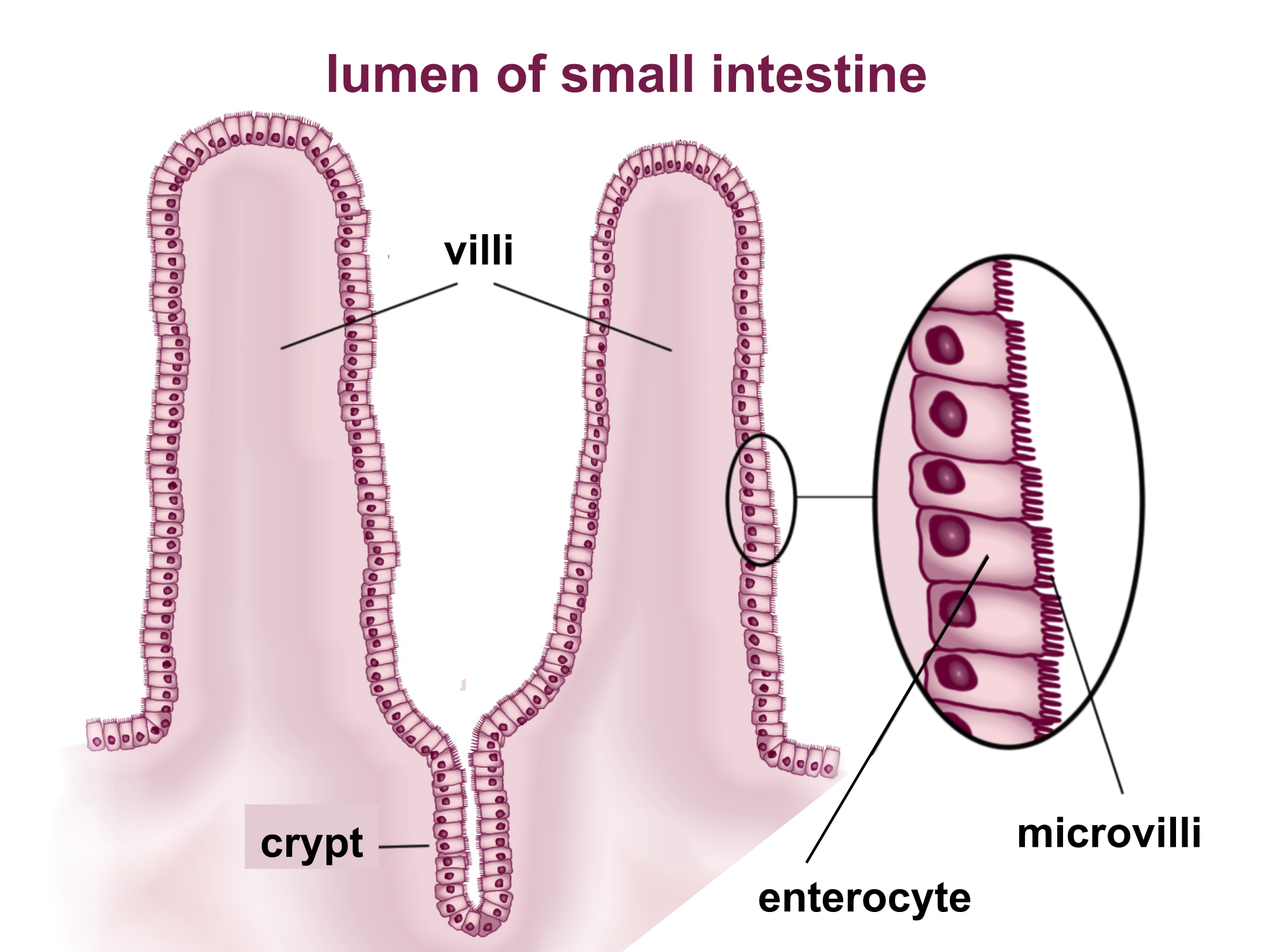
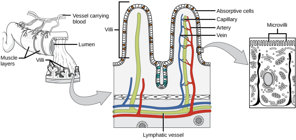
The small intestine is a narrow, but very long tubular organ. It may be almost seven metres long in adults. It is the site of most chemical digestion and virtually all absorption of nutrients. Many digestive enzymes are active in the small intestine, some of which are produced by the small intestine itself, and some of which are produced by the pancreas, an accessory organ of the digestive system. Much of the inner lining of the small intestine is covered by tiny finger-like projections called villi, each of which is covered by even tinier projections called microvilli. These projections, shown in the drawing below (Figure 13.6), greatly increase the surface area through which nutrients can be absorbed from the small intestine.
From the small intestine, any remaining nutrients and food waste pass into the large intestine. The large intestine is another tubular organ, but it is wider and shorter than the small intestine. It connects the small intestine and the anus. Waste that enters the large intestine is in a liquid state. As it passes through the large intestine, excess water is absorbed from it. The remaining solid waste — called feces — is eventually eliminated from the body through the anus.
ACCESSORY ORGANS OF THE DIGESTIVE SYSTEM

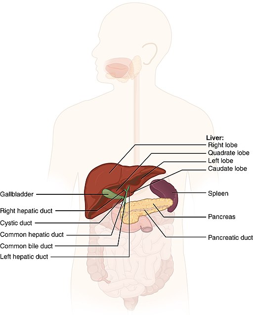
Accessory organs of the digestive system are not part of the GI tract, so they are not sites where digestion or absorption take place. Instead, these organs secrete or store substances needed for the chemical digestion of food. The accessory organs include the liver, gallbladder, and pancreas. They are shown in Figure 13.7 and described in the text that follows.
- The liver is an organ with multitude of functions. Its main digestive function is producing and secreting a fluid called bile, which reaches the small intestine through a duct. Bile breaks down large globules of lipids into smaller ones that are easier for enzymes to chemically digest. Bile is also needed to reduce the acidity of food entering the small intestine from the highly acidic stomach, because enzymes in the small intestine require a less acidic environment in order to work.
- The gallbladder is a small sac below the liver that stores some of the bile from the liver. The gallbladder also concentrates the bile by removing some of the water from it. It then secretes the concentrated bile into the small intestine as needed for fat digestion following a meal.
- The pancreas secretes many digestive enzymes and releases them into the small intestine for the chemical digestion of carbohydrates, proteins, and lipids. The pancreas also helps lessen the acidity of the small intestine by secreting bicarbonate, a basic substance that neutralizes acid.
Review
- What is the digestive system?
- What are the three main functions of the digestive system? Define each function.
- Relate the tissues in the walls of GI tract organs to the functions the organs perform.
13.3 UPPER GASTROINTESTINAL TRACT

Head Stand
Did you ever wonder what would happen if you tried to swallow food while standing on your head like this person in Figure 13.8? Many people think that food travels down the gullet from the mouth by the force of gravity. If that were the case, then food you swallowed would stay in your throat while you were standing on your head. In reality, your position doesn’t have much to do with your ability to swallow. Food will travel from your mouth to your stomach whether you are standing upright or upside down. That’s because the tube the food travels through — the esophagus — moves the food along via muscular contractions known as peristalsis. The esophagus is one of several organs that make up the upper gastrointestinal tract.
ORGANS OF THE UPPER GASTROINTESTINAL TRACT
Besides the esophagus, organs of the upper gastrointestinal (GI) tract include the mouth, pharynx, and stomach. These hollow organs are all connected to form a tube through which food passes during digestion. The only role in digestion played by the pharynx and esophagus is to move food through the GI tract. The mouth and stomach, in contrast, are organs where digestion — or the breakdown of food — also occurs. In both of these organs, food is broken into smaller pieces (mechanical digestion), as well as broken down chemically (chemical digestion). It should be noted that the first part of the small intestine (duodenum) is considered in some contexts to be part of the upper GI tract, but that practice is not followed here.
MOUTH
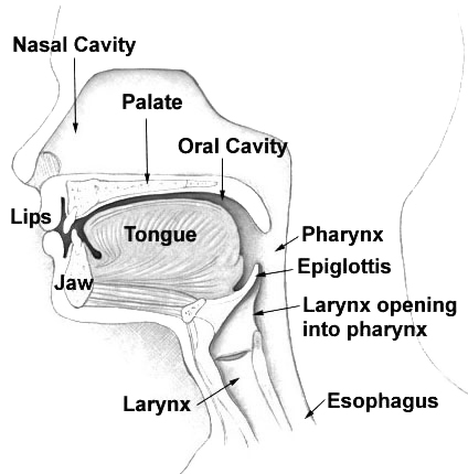
The mouth is the first organ of the GI tract. Most of the oral cavity is lined with mucous membrane. This tissue produces mucus, which helps moisten, soften, and lubricate food. Underlying the mucous membrane is a thin layer of smooth muscle to which the mucous membrane is only loosely connected. This gives the mucous membrane considerable ability to stretch as you eat food. The roof of the mouth, called the palate, separates the oral cavity from the nasal cavity. The front part is hard, consisting of mucous membrane covering a plate of bone. The back part of the palate is softer and more pliable, consisting of mucous membrane over muscle and connective tissue. The hard surface of the front of the palate allows for pressure needed in chewing and mixing food. The soft, pliable surface of the back of the palate can move to accommodate the passage of food while swallowing. Muscles at either side of the soft palate contract to create the swallowing action.
Several specific structures in the mouth are specialized for digestion. These include salivary glands, tongue, and teeth.
SALIVARY GLANDS
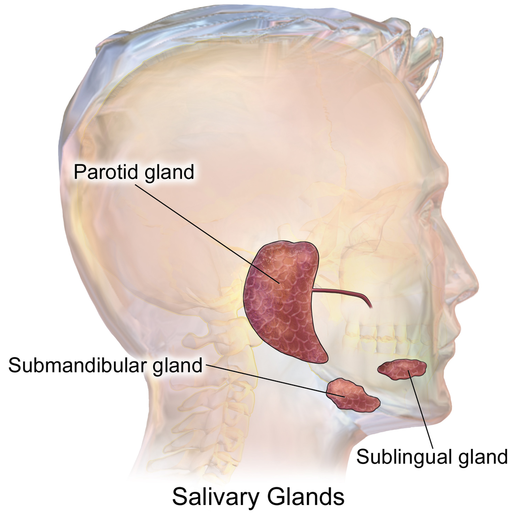
The mouth contains three pairs of major salivary glands, shown in Figure 13.9. These three pairs are all exocrine glands that secrete saliva into the mouth through ducts.
- The largest of the three major pairs of salivary glands are the parotid glands, which are located on either side of the mouth in front of the ears.
- The next largest pair is the submandibular glands, located beneath the lower jaw.
- The third pair is the sublingual glands, located underneath the tongue.
In addition to these three pairs of major salivary glands, there are also hundreds of minor salivary glands in the oral mucosa lining the mouth and on the tongue. Along with the major glands, most of the minor glands secrete the digestive enzyme amylase, which begins the chemical digestion of starch and glycogen (polysaccharides). However, the minor salivary glands on the tongue secrete the fat-digesting enzyme lipase, which in the mouth is called lingual lipase (to distinguish it from pancreatic lipase secreted by the pancreas).
Saliva secreted by the salivary glands mainly helps digestion, but it also plays other roles. It helps maintain dental health by cleaning the teeth, and it contains antibodies that help protect against infection. By keeping the mouth lubricated, saliva also allows the mouth movements needed for speech.
TONGUE
The tongue is a fleshy, muscular organ that is attached to the floor of the mouth by a band of ligaments that gives it great mobility. This is necessary so the tongue can manipulate food for chewing and swallowing. Movements of the tongue are also necessary for speaking. The upper surface of the tongue is covered with tiny projections called papillae, which contain taste buds. The latter are collections of chemoreceptor cells (shown in Figure 13.10). These sensory cells sense chemicals in food and send the information to the brain via cranial nerves, thus enabling the sense of taste.
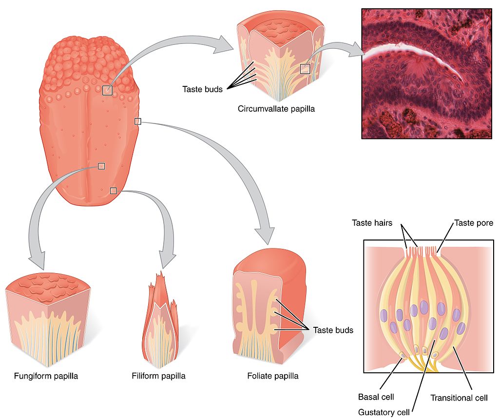
There are five basic tastes detected by the chemoreceptor cells in taste buds: saltiness, sourness, bitterness, sweetness, and umami (often described as a meaty taste). Contrary to popular belief, taste buds for the five basic tastes are not located on different parts of the tongue. Why does taste matter? The taste of food helps to stimulate the secretion of saliva from the salivary glands. It also helps us to eat foods that are good for us, instead of rotten or toxic foods. The detection of saltiness, for example, enables the control of salt intake and salt balance in the body. The detection of sourness may help us avoid spoiled foods, which often taste sour due to fermentation by bacteria. The detection of bitterness warns of poisons, because many plants defend themselves with toxins that taste bitter. The detection of sweetness guides us to foods that supply quick energy. The detection of umami may signal protein-rich foods.
TEETH
The teeth are complex structures made of a bone-like material called dentin and covered with enamel, which is the hardest tissue in the body. Adults normally have a total of 32 teeth, with 16 in each jaw. The right and left sides of each jaw are mirror images in terms of the numbers and types of teeth they contain. Teeth have different shapes to suit them for different aspects of mastication (chewing). The different types of teeth are illustrated in Figure 13.11.
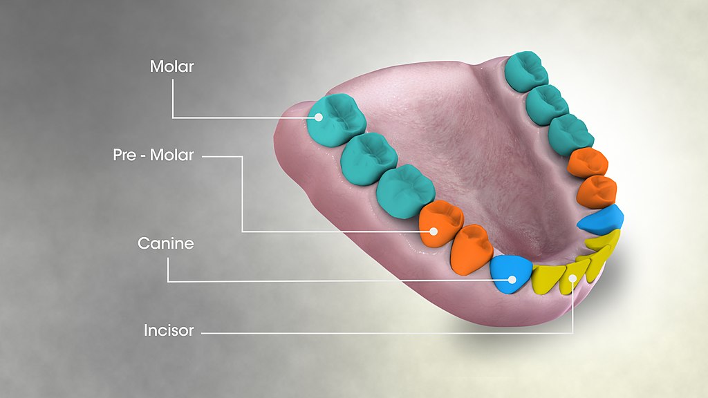
- Incisors are the sharp, blade-like teeth at the front of the mouth. They are used for cutting or biting off pieces of food. In adults, there are normally four incisors in each jaw, or eight in total.
- Canines are the pointed teeth on either side of the incisors. They are used for tearing foods that are tough or stringy. Adults normally have two canines in each jaw, or four altogether.
- Premolars and molars are cuboid teeth with cusps and grooves that are located on the sides and toward the back of the jaws. Premolars are closer to the front of the mouth. Molars are larger and have more cusps than premolars, but both are used for crushing and grinding food. Adults normally have two premolars and three molars on each side of each jaw, for a total of eight premolars and twelve molars.
PHARYNX
The tube-like pharynx (see Figure 13.12 below) plays a dual role as an organ of both respiration and digestion. As part of the respiratory system, it conducts air between the nasal cavity and larynx. As part of the digestive system, it allows swallowed food to pass from the oral cavity to the esophagus. Anything swallowed has priority over inhaled air when passing through the pharynx. During swallowing, the backward motion of the tongue causes a flap of elastic cartilage — called the epiglottis — to close over the opening to the larynx. This prevents food or drink from entering the larynx.
Esophagus
The esophagus (shown in Figure 13.13) is a muscular tube through which food is pushed from the pharynx to the stomach. The esophagus passes through an opening in the diaphragm (the large breathing muscle that separates the abdomen from the thorax) before reaching the stomach. In adults, the esophagus averages about 25 cm (about 9.8 inches) in length, depending on a person’s height. The inner lining of the esophagus consists of mucous membrane, which provides a smooth, slippery surface for the passage of food. The cells of this membrane are constantly being replaced as they are worn away from the frequent passage of food over them.
When food is not being swallowed, the esophagus is closed at both ends by upper and lower esophageal sphincters. Sphincters are rings of muscle that can contract to close off openings between structures. The upper esophageal sphincter is triggered to relax and open by the act of swallowing, allowing a bolus of food to enter the esophagus from the pharynx. Then, the esophageal sphincter closes again to prevent food from moving back into the pharynx from the esophagus.
Once in the esophagus, the food bolus travels down to the stomach, pushed along by the rhythmic contraction and relaxation of muscles (peristalsis). The lower esophageal sphincter is located at the junction between the esophagus and the stomach. This sphincter opens when the bolus reaches it, allowing the food to enter the stomach. The sphincter normally remains closed at other times to prevent the contents of the stomach from entering the esophagus. Failure of this sphincter to remain completely closed can lead to heartburn. If it happens chronically, it can lead to gastroesophageal reflux disease (GERD), in which the mucous membrane of the esophagus may become damaged by the highly acidic contents of the stomach.
See the video below to see how the parts of the upper GI tract work together to carry out swallowing: Swallowing, uploaded by Alejandra Cork, 2012.
Stomach
The stomach is a J-shaped organ (shown in Figure 13.14) that is joined to the esophagus at its upper end, and to the first part of the small intestine (duodenum) at its lower end. When the stomach is empty of food, it normally has a volume of about 75 millilitres, but it can expand to hold up to about a litre of food. Waves of muscle contractions (peristalsis) passing through the muscular walls of the stomach cause the food inside to be mixed and churned. The wall of the stomach has an extra layer of muscle tissue not found in other organs of the GI tract that helps it squeeze and mix the food. These movements of the stomach wall contribute greatly to mechanical digestion by breaking the food into much smaller pieces. The churning also helps mix the food with stomach secretions that aid in its chemical digestion.
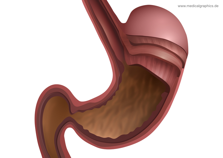
Secretions of the stomach include gastric acid, which consists mainly of hydrochloric acid (HCl). This makes the stomach contents highly acidic, which is necessary so that the enzyme pepsin — also secreted by the stomach — can begin the digestion of protein. Mucus is secreted by the lining of stomach to provide a slimy protective coating against the otherwise damaging effects of gastric acid. The fat-digesting enzyme lipase is secreted in small amounts in the stomach, but very little fat digestion occurs there.
By the time food has been in the stomach for about an hour, it has become the thick, semi-liquid chyme. When the small intestine is ready to receive chyme, a sphincter between the stomach and duodenum — called the pyloric sphincter — opens to allow the chyme to enter the small intestine for further digestion and absorption.
Review
- Identify structures in the mouth that are specialized for digestion.
- Describe digestion in the mouth.
- What general role do the pharynx and esophagus play in the digestion of food?
- How does food travel through the esophagus?
- Describe digestion in the stomach.
- Describe the differences between how air and food normally move past the pharynx.
- Name two structures in the mouth that contribute to mechanical digestion.
- What structure normally keeps stomach contents from backing up into the esophagus?
- Thirty minutes after you eat a meal, where is most of your food located? Explain your answer.
- What are two roles of mucus in the upper GI tract?
13.4 LOWER GASTROINTESTINAL TRACT
ORGANS OF THE LOWER GASTROINTESTINAL TRACT
Most of the bacteria that normally live in the lower gastrointestinal (GI) tract live in the large intestine. They have important and mutually beneficial relationships with the human organism. We provide them with a great place to live, and they provide us with many benefits, some of which you can read about below. Besides the large intestine and its complement of helpful bacteria, the lower GI tract also includes the small intestine. The latter is arguably the most important organ of the digestive system. It is where most chemical digestion and virtually all absorption of nutrients take place.
SMALL INTESTINE
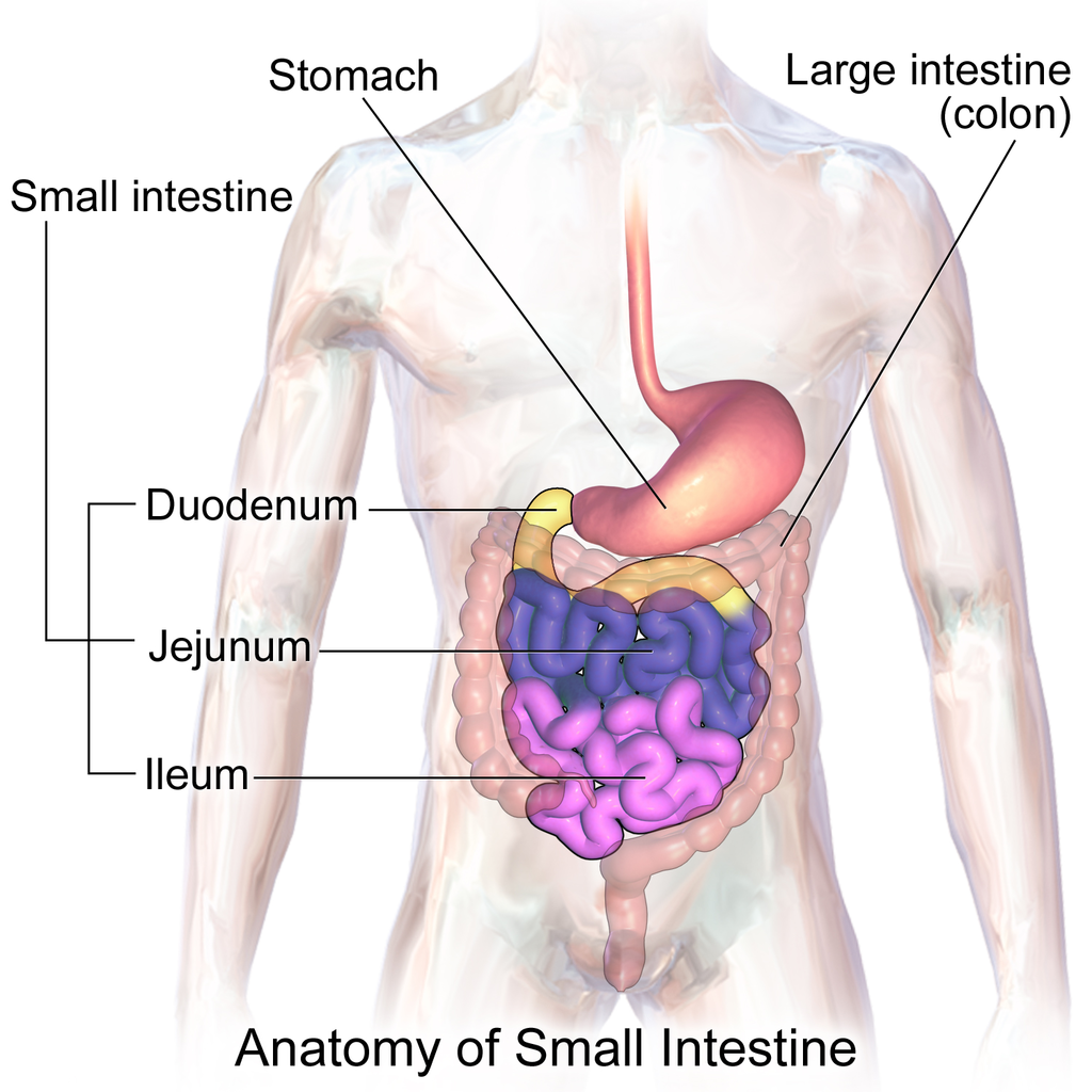
The small intestine (also called the small bowel or gut) is the part of the GI tract between the stomach and large intestine. Its average length in adults is 4.6 m in females and 6.9 m in males. It is approximately 2.5 to 3.0 cm in diameter. It is called “small” because it is much smaller in diameter than the large intestine. The internal surface area of the small intestine totals an average of about 30 m2. Structurally and functionally, the small intestine can be divided into three parts, called the duodenum, jejunum, and ileum, as shown in Figure 13.15 and described below.
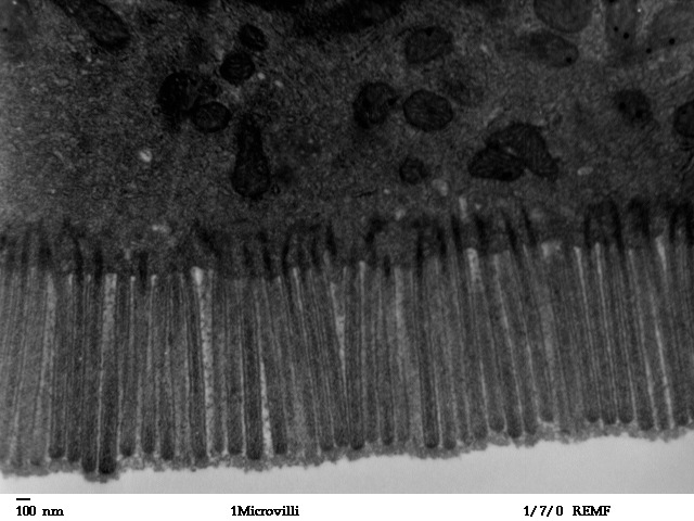
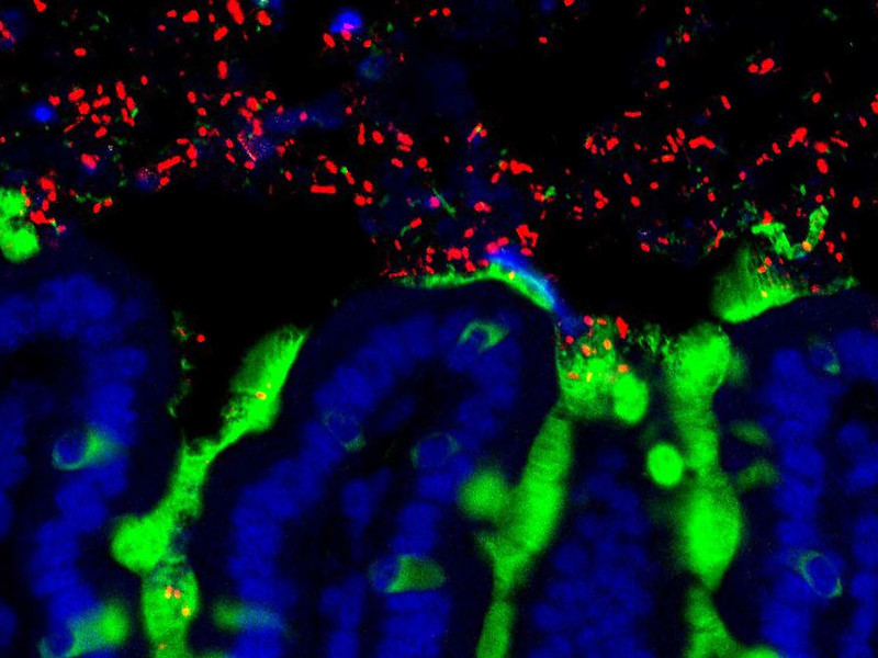
The mucosa lining the small intestine is highly enfolded and covered with the finger-like projections called villi. In fact, each square inch (about 6.5 cm2) of mucosa contains around 20 thousand villi. The individual cells on the surface of the villi also have many finger-like projections — the microvilli, shown in Figure 13.16. There are thought to be well over 17 billion microvilli per square centimetre of intestinal mucosa! All of these folds, villi, and microvilli greatly increase the surface area for chyme to come into contact with digestive enzymes, which coat the microvilli, as well as forming a tremendous surface area for the absorption of nutrients. Inside each of the villi is a network of tiny blood and lymph vessels that receive the absorbed nutrients and carry them away in the blood or lymph circulation. The wrinkles and projections in the intestinal mucosa also slow down the passage of chyme so there is more time for digestion and absorption to take place.
DUODENUM
The duodenum is the first part of the small intestine, directly connected to the stomach. It is also the shortest part of the small intestine, averaging only about 25 cm (almost 10 in) in length in adults. Its main function is chemical digestion, and it is where most of the chemical digestion in the entire GI tract takes place.
The duodenum receives partially digested, semi-liquid chyme from the stomach. It receives digestive enzymes and alkaline bicarbonate from the pancreas through the pancreatic duct, and it receives bile from the liver via the gallbladder through the common bile duct (see Figure 13.17). In addition, the lining of the duodenum secretes digestive enzymes and contains glands — called Brunner’s glands — that secrete mucous and bicarbonate. The bicarbonate from the pancreas and Brunner’s glands — along with bile from the liver — neutralize the highly acidic chyme after it enters the duodenum from the stomach. This is necessary because the digestive enzymes in the duodenum require a nearly neutral environment in order to work. The three major classes of compounds that undergo chemical digestion in the duodenum are carbohydrates, proteins, and lipids.
Digestion of Carbohydrates in the Duodenum
Complex carbohydrates (such as starches) are broken down by the digestive enzyme amylase from the pancreas to short-chain molecules consisting of just a few saccharides (that is, simple sugars). Disaccharides, including sucrose and lactose, are broken down to simple sugars by duodenal enzymes. Sucrase breaks down sucrose, and lactase (if present) breaks down lactose. Some carbohydrates are not digested in the duodenum, and they ultimately pass undigested to the large intestine, where they may be digested by intestinal bacteria.
Digestion of Proteins in the Duodenum
In the duodenum, the pancreatic enzymes trypsin and chymotrypsin cleave proteins into peptides. Then, these smaller molecules are broken down into amino acids. Their digestion is catalyzed by pancreatic enzymes called peptidases.
Digestion of Lipids in the Duodenum
Pancreatic lipase breaks down triglycerides into fatty acids and glycerol. Lipase works with the help of bile secreted by the liver and stored in the gallbladder. Bile salts attach to triglycerides to help them emulsify, or form smaller particles (called micelles) that can disperse through the watery contents of the duodenum. This increases access to the molecules by pancreatic lipase.
JEJUNUM
The jejunum is the middle part of the small intestine, connecting the duodenum and the ileum. The jejunum is about 2.5 m (about 8 ft) long. Its main function is absorption of the products of digestion, including sugars, amino acids, and fatty acids. Absorption occurs by simple diffusion (water and fatty acids), facilitated diffusion (the simple sugar fructose), or active transport (amino acids, small peptides, water-soluble vitamins, and most glucose). All nutrients are absorbed into the blood, except for fatty acids and fat-soluble vitamins, which are absorbed into the lymph. Although most nutrients are absorbed in the jejunum, there are a few exceptions:
- Iron is absorbed in the duodenum.
- Vitamin B12 and bile salts are absorbed in the ileum.
- Water and lipids are absorbed throughout the small intestine, including the duodenum and ileum in addition to the jejunum.
ILEUM
The ileum is the third and final part of the small intestine, directly connected at its distal end to the large intestine. The ileum is about 3 m (almost 10 ft) long. Some cells in the lining of the ileum secrete enzymes that catalyze the final stages of digestion of any undigested protein and carbohydrate molecules. However, the main function of the ileum is to absorb vitamin B12 and bile salts. It also absorbs any other remaining nutrients that were not absorbed in the jejunum. All substances in chyme that remain undigested or unabsorbed by the time they reach the distal end of the ileum pass into the large intestine.
LARGE INTESTINE
The large intestine— also called the large bowel — is the last organ of the GI tract. In adults, it averages about 1.5 m (about 5ft) in length. It is shorter than the small intestine, but at least twice as wide, averaging about 6.5 cm (about 2.5 in) in diameter. Water is absorbed from chyme as it passes through the large intestine, turning the chyme into solid feces. Feces is stored in the large intestine until it leaves the body during defecation.
PARTS OF THE LARGE INTESTINE
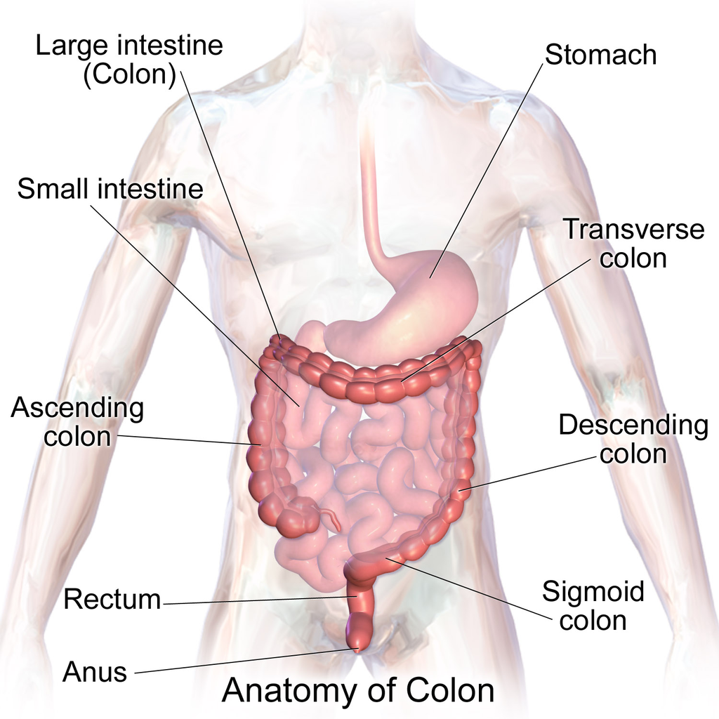
Like the small intestine, the large intestine can be divided into several parts, as shown in Figure 13.18. The large intestine begins at the end of the small intestine, where a valve separates the small and large intestines and regulates the movement of chyme into the large intestine. Most of the large intestine is called the colon. The first part of the colon, where chyme enters from the small intestine, is called the cecum. From the cecum, the colon continues upward as the ascending colon, travels across the upper abdomen as the transverse colon, and then continues downward as the descending colon. It then becomes a V-shaped region called the sigmoid colon, which is attached to the rectum. The rectum stores feces until elimination occurs. It transitions to the final part of the large intestine, called the anus, which has an opening to the outside of the body for feces to pass through.
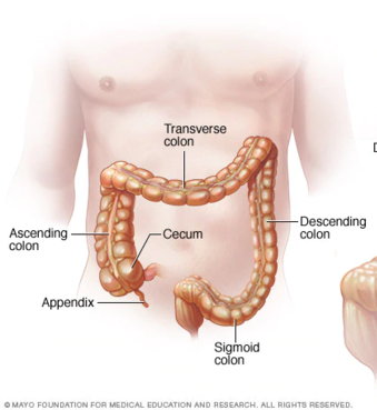
The diagram below (Figure 13.19) shows a projection from the cecum of the colon that is known as the appendix. The function of the appendix is somewhat uncertain, but it does not seem to be involved in digestion or absorption. It may play a role in immunity, and in the fetus, it seems to have an endocrine function, releasing hormones needed for homeostasis. Some biologists speculate that the appendix may also store a sample of the colon’s normal bacteria. If so, it may be able to repopulate the colon with the bacteria if illness or antibiotic medications deplete these microorganisms. Appendicitis, or infection and inflammation of the appendix, is a fairly common medical problem, typically resolved by surgical removal of the appendix (appendectomy). People who have had their appendix surgically removed do not seem to suffer any ill effects, so the organ is considered dispensable.
FUNCTIONS OF THE LARGE INTESTINE
The removal of water from chyme to form feces starts in the ascending colon and continues throughout much of the length of the organ. Salts (such as sodium) are also removed from food wastes in the large intestine before the wastes are eliminated from the body. This allows salts — as well as water — to be recycled in the body.
The large intestine is also the site where huge numbers of beneficial bacteria ferment many unabsorbed materials in food waste. The bacterial breakdown of undigested polysaccharides produces nitrogen, carbon dioxide, methane, and other gases responsible for intestinal gas, or flatulence. These bacteria are particularly prevalent in the descending colon. Some of the bacteria also produce vitamins that are absorbed from the colon. The vitamins include vitamins B1 (thiamine), B2 (riboflavin), B7 (biotin), B12, and K. Another role of bacteria in the colon is an immune function. The bacteria may stimulate the immune system to produce antibodies that are effective against similar, but pathogenic, bacteria, thereby preventing infections. Still other roles played by bacteria in the large intestine include breaking down toxins before they can poison the body, producing substances that help prevent colon cancer, and inhibiting the growth of harmful bacteria.
FEATURE: MY HUMAN BODY
Serotonin is a neurotransmitter with a wide variety of functions in the body. Sometimes called the “happy chemical,” it is used in the central nervous system to stabilize mood by contributing to a feeling of well being and happiness. While this “happy chemical” function is important, serotonin is also important for critical brain functions, including support of learning, memory, and reward structures and regulating sleep. Since serotonin’s main target is brain function, you may be surprised to learn that 90% of the human body’s supply of this neurotransmitter is located in the gastrointestinal tract, where it regulates intestinal movements.
The enteric nervous system (ENS), a division of the autonomic nervous system, is a mesh-like system of neurons that control GI function. Interestingly, the ENS can act independently from both the sympathetic/parasympathetic nervous systems and the brain/spinal cord, which is why some scientists refer to the ENS as the “second brain”. The ENS consists of over 500 million neurons (five times as many as in the spinal cord!) and lines the GI tract from the esophagus all the way to the anus. Serotonin is made in and secreted by the gut (small and large intestine) by enterochromaffin cells (also known as Kulchitsky cells) residing in the inner lining of the lower GI tract. While the main function of serotonin in the gut is to regulate digestion, it is also suspected to play a role in brain function — meaning that your gut may actually be partly dictating your mood! Another piece of evidence for the gut-dictating-your-mood theory is the nature vagus nerve, a fibrous visceral nerve, in which 90% of the fibers are dedicated to sending information to the brain, and only 10% of the fibers receiving information from the brain. Some treatments for depression actually involved electrical stimulation of the vagus nerve!
In a 2015 study by researchers at Caltech, it was shown that gut bacteria promote serotonin production by enterochromaffin cells. The study involved measuring the levels of serotonin levels in mice with normal gut bacteria, and then comparing that to the levels of serotonin in a population of mice with no gut bacteria. The germ-free mice were found to produce only 40% of the serotonin that the mice with normal gut flora were producing. Once the germ-free mice were re-colonized with normal gut flora, their production of serotonin returned to normal. This led researchers to the conclusion that enterochromaffin cells depend on an interaction with gut bacterial flora to be able to produce necessary levels of serotonin.
Review
- How is the mucosa of the small intestine specialized for digestion and absorption?
- What digestive substances are secreted into the duodenum? What compounds in food do they help digest?
- What is the main function of the jejunum?
- What roles does the ileum play?
- How do beneficial bacteria in the large intestine help the human organism?
- When diarrhea occurs, feces leaves the body in a more liquid state than normal. What part of the digestive system do you think is involved in diarrhea? Explain your answer
- What causes intestinal gas, or flatulence?
What causes constipation? – Heba Shaheed, TED-Ed, 2018.
13.5 ACCESSORY ORGANS OF DIGESTION

JAUNDICED EYES
Did you ever hear of a person looking at something or someone with a “jaundiced eye”? It means to take a negative view, such as envy, maliciousness, or ill will. The expression may be based on the antiquated idea that liver bile is associated with such negative emotions as these, as well as the fact that excessive liver bile causes jaundice, or yellowing of the eyes and skin. Jaundice is likely a sign of a liver disorder or blockage of the duct that carries bile away from the liver. Bile contains waste products, making the liver an organ of excretion. Bile has an important role in digestion, which makes the liver an accessory organ of digestion, too.
WHAT ARE ACCESSORY ORGANS OF DIGESTION?
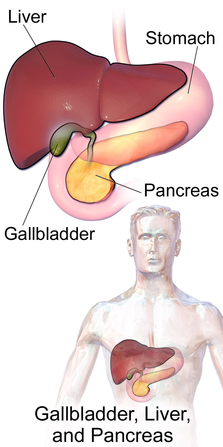
Accessory organs of digestion are organs that secrete substances needed for the chemical digestion of food, but through which food does not actually pass as it is digested. Besides the liver, the major accessory organs of digestion are the gallbladder and pancreas. These organs secrete or store substances that are needed for digestion in the first part of the small intestine — the duodenum — where most chemical digestion takes place. You can see the three organs and their locations in Figure 13.21.
Liver
The liver is a vital organ located in the upper right part of the abdomen. It lies just below the diaphragm, to the right of the stomach. The liver plays an important role in digestion by secreting bile, but the liver has a wide range of additional functions unrelated to digestion. In fact, some estimates put the number of functions of the liver at about 500! A few of them are described below.
STRUCTURE OF THE LIVER
The liver is a reddish brown, wedge-shaped structure. In adults, the liver normally weighs about 1.5 kg (about 3.3 lb.). It is both the heaviest internal organ and the largest gland in the human body. The liver is divided into four lobes of unequal size and shape. Each lobe, in turn, is made up of lobules, which are the functional units of the liver. Each lobule consists of millions of liver cells, called hepatic cells (or hepatocytes). They are the basic metabolic cells that carry out the various functions of the liver.
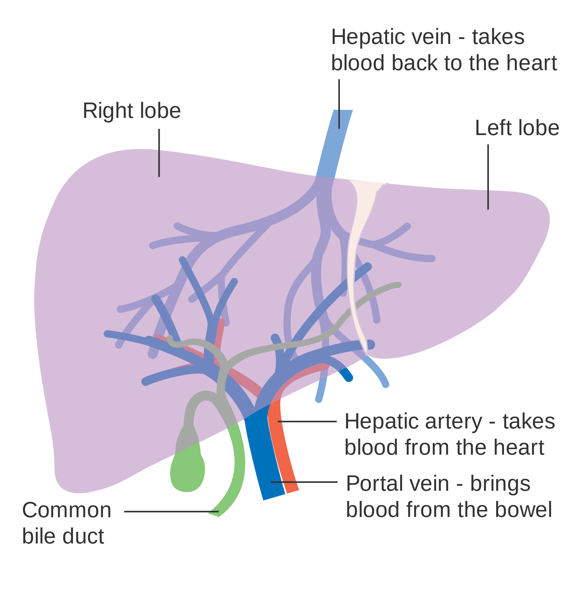
As shown in Figure 13.22, the liver is connected to two large blood vessels: the hepatic artery and the portal vein. The hepatic artery carries oxygen-rich blood from the aorta, whereas the portal vein carries blood that is rich in digested nutrients from the GI tract and wastes filtered from the blood by the spleen. The blood vessels subdivide into smaller arteries and capillaries, which lead into the liver lobules. The nutrients from the GI tract are used to build many vital biochemical compounds, and the wastes from the spleen are degraded and excreted.
FUNCTIONS OF THE LIVER
The main digestive function of the liver is the production of bile. Bile is a yellowish alkaline liquid that consists of water, electrolytes, bile salts, and cholesterol, among other substances, many of which are waste products. Some of the components of bile are synthesized by hepatocytes. The rest are extracted from the blood.
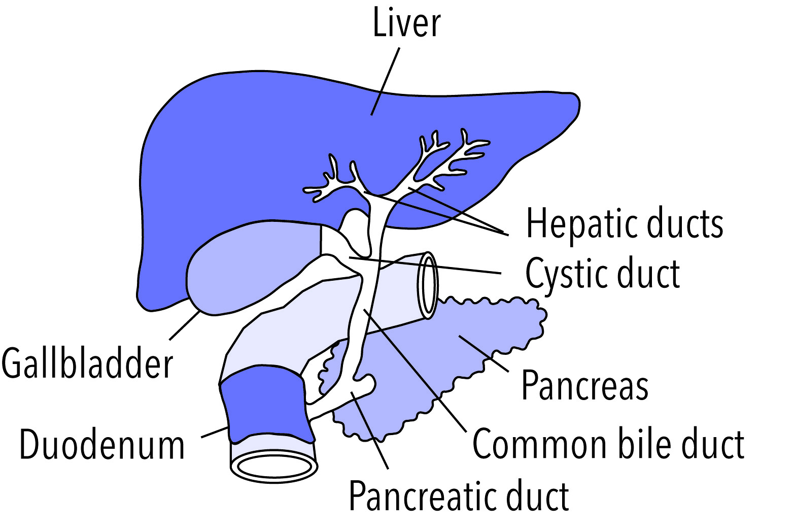
As shown in Figure 13.23, bile is secreted into small ducts that join together to form larger ducts, with just one large duct carrying bile out of the liver. If bile is needed to digest a meal, it goes directly to the duodenum through the common bile duct. In the duodenum, the bile neutralizes acidic chyme from the stomach and emulsifies fat globules into smaller particles (called micelles) that are easier to digest chemically by the enzyme lipase. Bile also aids with the absorption of vitamin K. Bile that is secreted when digestion is not taking place goes to the gallbladder for storage until the next meal. In either case, the bile enters the duodenum through the common bile duct.
Besides its roles in digestion, the liver has many other vital functions:
- The liver synthesizes glycogen from glucose and stores the glycogen as required to help regulate blood sugar levels. It also breaks down the stored glycogen to glucose and releases it back into the blood as needed.
- The liver stores many substances in addition to glycogen, including vitamins A, D, B12, and K. It also stores the minerals iron and copper.
- The liver synthesizes numerous proteins and many of the amino acids needed to make them. These proteins have a wide range of functions. They include fibrinogen, which is needed for blood clotting; insulin-like growth factor (IGF-1), which is important for childhood growth; and albumen, which is the most abundant protein in blood serum and functions to transport fatty acids and steroid hormones in the blood.
- The liver synthesizes many important lipids, including cholesterol, triglycerides, and lipoproteins.
- The liver is responsible for the breakdown of many waste products and toxic substances. The wastes are excreted in bile or travel to the kidneys, which excrete them in urine.
The liver is clearly a vital organ that supports almost every other organ in the body. Because of its strategic location and diversity of functions, the liver is also prone to many diseases, some of which cause loss of liver function. There is currently no way to compensate for the absence of liver function in the long term, although liver dialysis techniques can be used in the short term. An artificial liver has not yet been developed, so liver transplantation may be the only option for people with liver failure.
GALLBLADDER
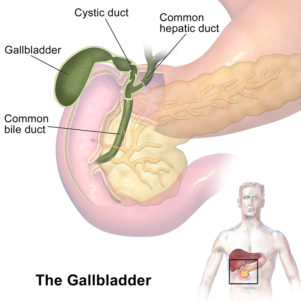
The gallbladder is a small, hollow, pouch-like organ that lies just under the right side of the liver (see Figure 13.24). It is about 8 cm (about 3 in) long and shaped like a tapered sac, with the open end continuous with the cystic duct. The gallbladder stores and concentrates bile from the liver until it is needed in the duodenum to help digest lipids. After the bile leaves the liver, it reaches the gallbladder through the cystic duct. At any given time, the gallbladder may store between 30 to 60 mL (1 to 2 oz) of bile. A hormone stimulated by the presence of fat in the duodenum signals the gallbladder to contract and force its contents back through the cystic duct and into the common bile duct to drain into the duodenum.
PANCREAS
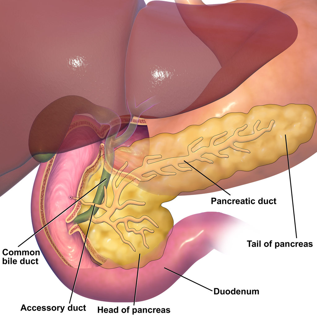
The pancreas is a glandular organ that is part of both the digestive system and the endocrine system. As shown in Figure 13.25, it is located in the abdomen behind the stomach, with the head of the pancreas surrounded by the duodenum of the small intestine. The pancreas is about 15 cm (almost 6 in) long, and it has two major ducts: the main pancreatic duct and the accessory pancreatic duct. Both of these ducts drain into the duodenum.
As an endocrine gland, the pancreas secretes several hormones, including insulin and glucagon, which circulate in the blood. The endocrine hormones are secreted by clusters of cells called pancreatic islets (or islets of Langerhans). As a digestive organ, the pancreas secretes many digestive enzymes and also bicarbonate, which helps neutralize acidic chyme after it enters the duodenum. The pancreas is stimulated to secrete its digestive substances when food in the stomach and duodenum triggers the release of endocrine hormones into the blood that reach the pancreas via the bloodstream. The pancreatic digestive enzymes are secreted by clusters of cells called acini, and they travel through the pancreatic ducts to the duodenum. In the duodenum, they help to chemically break down carbohydrates, proteins, lipids, and nucleic acids in chyme. The pancreatic digestive enzymes include:
- Amylase, which helps digest starch and other carbohydrates.
- Trypsin and chymotrypsin, which help digest proteins.
- Lipase, which helps digest lipids.
- Deoxyribonucleases and ribonucleases, which help digest nucleic acids.
Review
- Name three accessory organs of digestion. How do these organs differ from digestive organs that are part of the GI tract?
-
Fill in the blanks:
The ____________ has many functions, some of which are producing bile, converting glucose to glycogen, and breakdown of toxic substances.
The ____________ stores bile and then contracts to force its contents into the duodenum when needed.
The ____________ is both an endocrine organ and a digestive organ.
The ____________ exits the gallbladder, the ____________ exit the liver, and the ____________ exits the pancreas. The first two ducts merge into the ______________ and then deliver their contents to the ____________ .
- Describe the liver and its blood supply.
- Explain the main digestive function of the liver and describe the components of bile and it’s importance in the digestive process.
- What type of secretions does the pancreas release as part of each body system?
- List pancreatic enzymes that work in the duodenum, along with the substances they help digest.
- What are two substances produced by accessory organs of digestion that help neutralize chyme in the small intestine? Where are they produced?
- People who have their gallbladder removed sometimes have digestive problems after eating high-fat meals. Why do you think this happens?
- Which accessory organ of digestion synthesizes cholesterol?
What does the pancreas do? – Emma Bryce, TED-Ed. 2015.
What does the liver do? – Emma Bryce, TED-Ed, 2014.
13.6 DIGESTION AND ABSORPTION
DIGESTION
Digestion of food is a form of catabolism, in which the food is broken down into small molecules that the body can absorb and use for energy, growth, and repair. Digestion occurs when food is moved through the digestive system. This process begins in the mouth and ends in the small intestine. The final products of digestion are absorbed from the digestive tract, primarily in the small intestine. There are two different types of digestion that occur in the digestive system: mechanical digestion and chemical digestion. Figure 13.26 summarizes the roles played by different digestive organs in mechanical and chemical digestion, both of which are described in detail below.
MECHANICAL DIGESTION

Mechanical digestion is a physical process in which food is broken into smaller pieces without becoming changed chemically. It begins with your first bite of food (see Figure 13.27) and continues as you chew food with your teeth into smaller pieces. The process of mechanical digestion continues in the stomach. This muscular organ churns and mixes the food it contains, an action that breaks any solid food into still smaller pieces.
Although some mechanical digestion also occurs in the small intestine, it is mostly completed by the time food leaves the stomach. At that stage, food in the GI tract has been changed to the thick semi-fluid called chyme. Mechanical digestion is necessary so that chemical digestion can be effective. Mechanical digestion tremendously increases the surface area of food particles so they can be acted upon more effectively by digestive enzymes.
CHEMICAL DIGESTION
Chemical digestion is the biochemical process in which macromolecules in food are changed into smaller molecules that can be absorbed into body fluids and transported to cells throughout the body. Substances in food that must be chemically digested include carbohydrates, proteins, lipids, and nucleic acids. Carbohydrates must be broken down into simple sugars, proteins into amino acids, lipids into fatty acids and glycerol, and nucleic acids into nitrogen bases and sugars. Some chemical digestion takes place in the mouth and stomach, but most of it occurs in the first part of the small intestine (duodenum).
Digestive Enzymes
Chemical digestion could not occur without the help of many different digestive enzymes. Enzymes are proteins that catalyze, or speed up, biochemical reactions. Digestive enzymes are secreted by exocrine glands or by the mucosal layer of epithelium lining the gastrointestinal tract. In the mouth, digestive enzymes are secreted by salivary glands. The lining of the stomach secretes enzymes, as does the lining of the small intestine. Many more digestive enzymes are secreted by exocrine cells in the pancreas and carried by ducts to the small intestine. The following table lists several important digestive enzymes, the organs and/or glands that secrete them, the compounds they digest, and the pH necessary for optimal functioning. You can read more about them below.
| Digestive Enzyme | Source Organ | Site of Action | Reactant and Product | Optimal pH |
|---|---|---|---|---|
| Salivary Amylase | Salivary Glands | Mouth | starch + water ⇒ maltose | Neutral |
| Pepsin | Stomach | Stomach | protein + water ⇒ peptides | Acidic |
| Pancreatic Amylase | Pancreas | Duodenum | starch + water ⇒ maltose | Basic |
| Maltase | Small intestine | Small intestine | maltose + water ⇒ glucose | Basic |
| Sucrase | Small intestine | Small intestine | sucrose + water ⇒ glucose + fructose | Basic |
| Lactase | Small intestine | Small intestine | lactose + water ⇒ glucose + galactose | Basic |
| Lipase | Pancreas | Duodenum | fat droplet and water ⇒ glycerol and fatty acids | Basic |
| Trypsin | Pancreas | Duodenum | protein + water ⇒ peptides | Basic |
| Chymotrypsin | Pancreas | Duodenum | protein + water ⇒ peptides | Basic |
| Peptidases | Small intestine | Small intestine | peptides + water ⇒ | Basic |
| Deoxyribonuclease | Pancreas | Duodenum | DNA + water ⇒ nucleotide fragments | Basic |
| Ribonuclease | Pancreas | Duodenum | RNA + water ⇒ nucleotide fragments | Basic |
| Nuclease | Small intestine | Small intestine | nucleic acids + water ⇒ nucleotide fragments | Basic |
| Nucleosidases | Small intestine | Small intestine | nucleotides + water ⇒ nitrogen base + phosphate sugar | Basic |
Chemical Digestion of Carbohydrates
About 80% of digestible carbohydrates in a typical Western diet are in the form of the plant polysaccharide amylose, which consists mainly of long chains of glucose and is one of two major components of starch. Additional dietary carbohydrates include the animal polysaccharide glycogen, along with some sugars, which are mainly disaccharides.
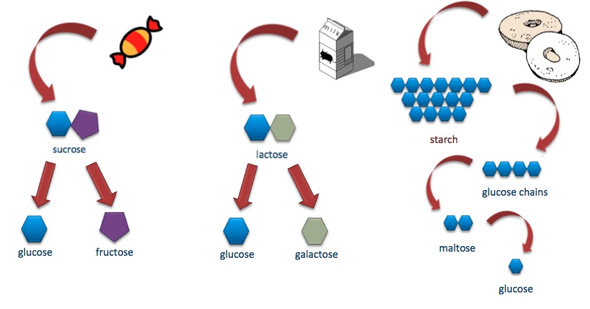
The process of chemical digestion for some carbohydrates is illustrated Figure 13.28. To chemically digest amylose and glycogen, the enzyme amylase is required. The chemical digestion of these polysaccharides begins in the mouth, aided by amylase in saliva. Saliva also contains mucus — which lubricates the food — and hydrogen carbonate, which provides the ideal alkaline conditions for amylase to work. Carbohydrate digestion is completed in the small intestine, with the help of amylase secreted by the pancreas. In the digestive process, polysaccharides are reduced in length by the breaking of bonds between glucose monomers. The macromolecules are broken down to shorter polysaccharides and disaccharides, resulting in progressively shorter chains of glucose. The end result is molecules of the simple sugars glucose and maltose (which consists of two glucose molecules), both of which can be absorbed by the small intestine.
Other sugars are digested with the help of different enzymes produced by the small intestine. Sucrose (or table sugar), for example, is a disaccharide that is broken down by the enzyme sucrase to form glucose and fructose, which are readily absorbed by the small intestine. Digestion of the sugar lactose, which is found in milk, requires the enzyme lactase, which breaks down lactose into glucose and galactose. Glucose and galactose are then absorbed by the small intestine. Fewer than half of all adults produce sufficient lactase to be able to digest lactose. Those who cannot are said to be lactose intolerant.
Chemical Digestion of Proteins
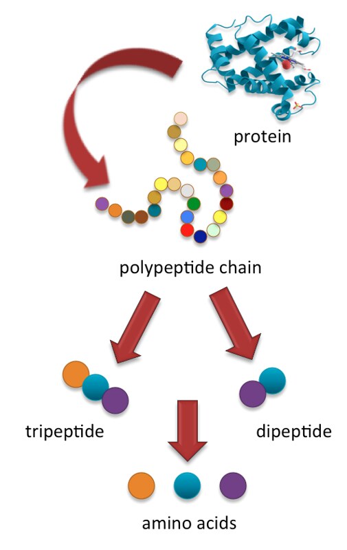
Proteins consist of polypeptides, which must be broken down into their constituent amino acids before they can be absorbed. An overview of this process is shown in Figure 13.29. Protein digestion occurs in the stomach and small intestine through the action of three primary enzymes: pepsin (secreted by the stomach), and trypsin and chymotrypsin (secreted by the pancreas). The stomach also secretes hydrochloric acid (HCl), making the contents highly acidic, which is a required condition for pepsin to work. Trypsin and chymotrypsin in the small intestine require an alkaline (basic) environment to work. Bile from the liver and bicarbonate from the pancreas neutralize the acidic chyme as it empties into the small intestine. After pepsin, trypsin, and chymotrypsin break down proteins into peptides, these are further broken down into amino acids by other enzymes called peptidases, also secreted by the pancreas.
Chemical Digestion of Lipids
The chemical digestion of lipids begins in the mouth. The salivary glands secrete the digestive enzyme lipase, which breaks down short-chain lipids into molecules consisting of two fatty acids. A tiny amount of lipid digestion may take place in the stomach, but most lipid digestion occurs in the small intestine.
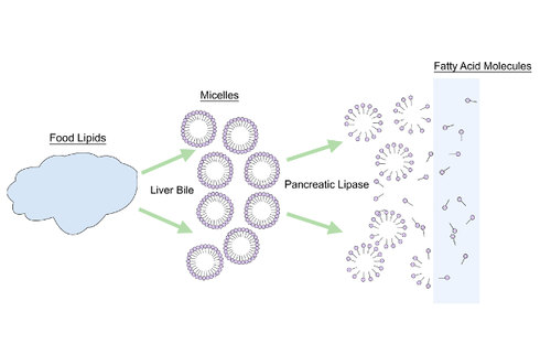
Digestion of lipids in the small intestine occurs with the help of another lipase enzyme from the pancreas, as well as bile secreted by the liver. As shown in the diagram below (Figure 13.30), bile is required for the digestion of lipids, because lipids are oily and do not dissolve in the watery chyme. Bile emulsifies (or breaks up) large globules of food lipids into much smaller ones, called micelles, much as dish detergent breaks up grease. The micelles provide a great deal more surface area to be acted upon by lipase, and also point the hydrophilic (“water-loving”) heads of the fatty acids outward into the watery chyme. Lipase can then access and break down the micelles into individual fatty acid molecules.
Chemical Digestion of Nucleic Acids
Nucleic acids (DNA and RNA) in foods are digested in the small intestine with the help of both pancreatic enzymes and enzymes produced by the small intestine itself. Pancreatic enzymes called ribonuclease and deoxyribonuclease break down RNA and DNA, respectively, into smaller nucleic acids. These, in turn, are further broken down into nitrogen bases and sugars by small intestine enzymes called nucleases.
Bacteria in the Digestive System
Your large intestine is not just made up of cells. It is also an ecosystem, home to trillions of bacteria known as the “gut flora” (Figure 13.30). But don’t worry, most of these bacteria are helpful. Friendly bacteria live mostly in the large intestine and part of the small intestine. The acidic environment of the stomach does not allow bacterial growth.
Gut bacteria have several roles in the body. For example, intestinal bacteria:
- Produce vitamin B12 and vitamin K.
- Control the growth of harmful bacteria.
- Break down poisons in the large intestine.
- Break down some substances in food that cannot be digested, such as fibre and some starches and sugars. Bacteria produce enzymes that digest carbohydrates in plant cell walls. Most of the nutritional value of plant material would be wasted without these bacteria. These help us digest plant foods like spinach.
A wide range of friendly bacteria live in the gut. Bacteria begin to populate the human digestive system right after birth. Gut bacteria include Lactobacillus, the bacteria commonly used in probiotic foods such as yogurt, and E. coli bacteria. About a third of all bacteria in the gut are members of the Bacteroides species. Bacteroides are key in helping us digest plant food.
It is estimated that 100 trillion bacteria live in the gut. This is more than the human cells that make up you. It has also been estimated that there are more bacteria in your mouth than people on the planet — there are over 7 billion people on the planet!
The bacteria in your digestive system are from anywhere between 300 and 1,000 species. As these bacteria are helpful, your body does not attack them. They actually appear to the body’s immune system as cells of the digestive system, not foreign invaders. The bacteria actually cover themselves with sugar molecules removed from the actual cells of the digestive system. This disguises the bacteria and protects them from the immune system.
As the bacteria that live in the human gut are beneficial to us, and as the bacteria enjoy a safe environment to live, the relationship that we have with these tiny organisms is described as mutualism, a type of symbiotic relationship.
Lastly, keep in mind the small size of bacteria. Together, all the bacteria in your gut may weigh just about two pounds.
CONTROL OF THE DIGESTIVE PROCESS
The process of digestion is controlled by both hormones and nerves. Hormonal control is mainly by endocrine hormones secreted by cells in the lining of the stomach and small intestine. These hormones stimulate the production of digestive enzymes, bicarbonate, and bile. The hormone secretin, for example, is produced by endocrine cells lining the duodenum of the small intestine. Acidic chyme entering the duodenum from the stomach triggers the release of secretin into the bloodstream. When the secretin returns via the circulation to the digestive system, it signals the release of bicarbonate from the pancreas. The bicarbonate neutralizes the acidic chyme. See Table 13.2 for a summary of the major hormones governing the process of chemical digestion.
| Hormone | Source Organ | Target Organ | Trigger | Result |
|---|---|---|---|---|
| Gastrin | Stomach walls | Stomach | High protein intake | HCL and pepsin release, stomach churning |
| Secretin | Duodenum | Pancreas
Gallbladder |
Acidic chyme entering the duodenum | Release sodium bicarbonate, release bile |
| Cholecystokinin (CCK) | Duodenum | Pancreas
Gallbladder |
Partially digested fat and protein in duodenum | Release lipase, trypsin, release bile |
Nerves involved in digestion include those that connect digestive organs to the central nervous system, as well as nerves inside the walls of the digestive organs. Nerves connecting the digestive organs to the central nervous system cause smooth muscles in the walls of digestive organs to contract or relax as needed, depending on whether or not there is food to be digested. Nerves within digestive organs are stimulated when food enters the organs and stretches their walls. These nerves trigger the release of substances that speed up or slow down the movement of food through the GI tract and the secretion of digestive enzymes.
ABSORPTION
When digestion is finished, it results in many simple nutrient molecules that must go through the process of absorption from the lumen of the GI tract to blood or lymph vessels, so they can be transported to and used by cells throughout the body. A few substances are absorbed in the stomach and large intestine. Water is absorbed in both of these organs, and some minerals and vitamins are also absorbed in the large intestine, but about 95% of nutrient molecules are absorbed in the small intestine. Absorption of the majority of these molecules takes place in the second part of the small intestine, called the jejunum. There are, however, a few exceptions — for example, iron is absorbed in the duodenum, and vitamin B12 is absorbed in the last part of the small intestine, called the ileum. After being absorbed in the small intestine, nutrient molecules are transported to other parts of the body for storage or further chemical modification. Amino acids, for instance, are transported to the liver to be used for protein synthesis.
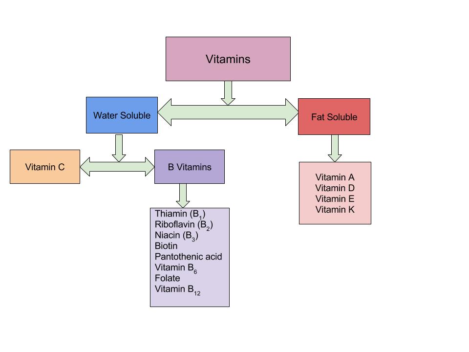
The epithelial tissue lining the small intestine is specialized for absorption. It is highly enfolded and is covered with villi and microvilli, creating an enormous surface area for absorption. As shown in Figure 13.32, each villus also has a network of blood capillaries and fine lymphatic vessels called lacteals close to its surface. The thin surface layer of epithelial cells of the villi transports nutrients from the lumen of the small intestine into these capillaries and lacteals. Blood in the capillaries absorbs most of the molecules, including simple sugars, amino acids, glycerol, salts, and water-soluble vitamins (vitamin C and the many B vitamins). Lymph in the lacteals absorbs fatty acids and fat-soluble vitamins (vitamins A, D, E, and K).
Feature: My Human Body
The process of digestion does not always go as it should. Many people suffer from indigestion, or dyspepsia, a condition of impaired digestion. Symptoms may include upper abdominal fullness or pain, heartburn, nausea, belching, or some combination of these symptoms. The majority of cases of indigestion occur without evidence of an organic disease that is likely to explain the symptoms. Anxiety or certain foods or medications (such as aspirin) may be contributing factors in these cases. In other cases, indigestion is a symptom of an organic disease, most often gastroesophageal reflux disease (GERD) or gastritis. In a small minority of cases, indigestion is a symptom of a peptic ulcer of the stomach or duodenum, usually caused by a bacterial infection. Very rarely, indigestion is a sign of cancer.
An occasional bout of indigestion is usually nothing to worry about, especially in people less than 55 years of age. However, if you suffer frequent or chronic indigestion, it’s a good idea to see a doctor. If an underlying disorder such as GERD or an ulcer is causing the indigestion, this can and should be treated. If no organic disease is discovered, the doctor can recommend lifestyle changes or treatments to help prevent or soothe the symptoms of acute indigestion. Lifestyle changes might include modifications in eating habits, such as eating more slowly, eating smaller meals, or avoiding fatty foods. You also might be advised to refrain from taking certain medications, especially on an empty stomach. The use of antacids or other medications to relieve symptoms may also be recommended.
Review
- Define digestion. Where does it occur?
-
What is the function of pepsin?
- Identify two organ systems that control the process of digestion by the digestive system.
- What is mechanical digestion? Where does it occur?
- Describe chemical digestion.
- What is the role of enzymes in chemical digestion?
- What is absorption? When does it occur?
- Where does most absorption occur in the digestive system? Why does most of the absorption occur in this organ, and not earlier in the GI tract?
13.7 NUTRITION
Nutrition is defined as the interaction of an organism and its food. There are several ways to get the recommended nutrition you need: a Nutritionist, your private physician, the United State Department of Agriculture (USDA) and the Department of Health and Human Services. It is important that you get the proper nutrition to maintain homeostasis in the body as you will read in the rest of this chapter.
The foods and beverages that people consume have a profound impact on their health. The scientific connection between food and health has been well documented for many decades, with substantial evidence showing that healthy dietary patterns can help people achieve and maintain good health and reduce the risk of chronic diseases throughout all stages of the lifespan. Yet, Federal data show that from the first edition of the Dietary Guidelines for Americans in 1980 through today, Americans have fallen far short of meeting its recommendations, and diet-related chronic disease rates have risen to pervasive levels and continue to be a major public health concern. The U.S. Departments of Agriculture (USDA) and of Health and Human Services (HHS) update the Dietary Guidelines at least every 5 years, based on the current science. A fundamental premise of the Dietary Guidelines is that everyone, no matter their age, race, or ethnicity, economic circumstances, or health status, can benefit from shifting food and beverage choices to better support healthy dietary patterns.
Major Themes of the Dietary Guidelines
Consume a healthy eating pattern that accounts for all foods and beverages within an appropriate calorie level. A healthy eating pattern includes:
- A variety of vegetables from all of the subgroups—dark green, red and orange, legumes (beans and peas), starchy, and other
- Fruits, especially whole fruits
- Grains, at least half of which are whole grains
- Fat-free or low-fat dairy, including milk, yogurt, cheese, and/or fortified soy beverages
- A variety of protein foods, including seafood, lean meats and poultry, eggs, legumes (beans and peas), and nuts, seeds, and soy products
Oils
A healthy eating pattern limits:
- Saturated fats and trans fats, added sugars, and sodium
- Cholesterol, in order to limit saturated fats.
Key Recommendations that are quantitative are provided for several components of the diet that should be limited. These components are of particular public health concern in the United States, and the specified limits can help individuals achieve healthy eating patterns within calorie limits:
- Consume less than 10 percent of calories per day from added sugars
- Consume less than 10 percent of calories per day from saturated fats
- Consume less than 2,300 milligrams (mg) per day of sodium
- Limit alcohol consumption —up to one drink per day for women and up to two drinks per day for men..
High consumptions of certain foods, such as those high in saturated or trans-fat, sodium, added sugars, and refined grains may contribute to the increased incidence of chronic disease. Additionally, excessive consumption of these foods replaces the intake of more nutrient-dense foods.
My Plate
Estimating portions can be done using the MyPlate Planner. Recall that the MyPlate symbol is divided according to how much of each food group should be included with each meal. Note the MyPlate Planner Methods of Use:
- Fill half of your plate with vegetables such as carrots, broccoli, salad, and fruit.
- Fill one-quarter of your plate with lean meat, chicken, or fish (about 3 ounces)
- Fill one-quarter of your plate with a whole grain such as ⅓ cup rice
- Choose one serving of dairy
- Add margarine or oil for preparation or addition at the table
For information about on My Plate go to the USDA’s site: MyPlate | U.S. Department of Agriculture
Figure 13.33 Choose My Plate
What are Nutrients?
The foods we eat contain nutrients. Nutrients are substances required by the body to perform its basic functions. Nutrients must be obtained from our diet, since the human body does not synthesize or produce them. Nutrients have one or more of three basic functions: they provide energy, contribute to body structure, and/or regulate chemical processes in the body. These basic functions allow us to detect and respond to environmental surroundings, move, excrete wastes, respire (breathe), grow, and reproduce. There are six classes of nutrients required for the body to function and maintain overall health. These are carbohydrates, lipids, proteins, water, vitamins, and minerals. Foods also contain non-nutrients that may be harmful (such as natural toxins common in plant foods and additives like some dyes and preservatives) or beneficial (such as antioxidants).
Macronutrients
Nutrients that are needed in large amounts are called macronutrients. There are three classes of macronutrients: carbohydrates, lipids, and proteins. These can be metabolically processed into cellular energy. The energy from macronutrients comes from their chemical bonds. This chemical energy is converted into cellular energy that is then utilized to perform work, allowing our bodies to conduct their basic functions. A unit of measurement of food energy is the calorie. On nutrition food labels the amount given for “calories” is actually equivalent to each calorie multiplied by one thousand. A kilocalorie (one thousand calories, denoted with a small “c”) is synonymous with the “Calorie” (with a capital “C”) on nutrition food labels. Water is also a macronutrient in the sense that you require a large amount of it, but unlike the other macronutrients, it does not yield calories.
Micronutrients
Micronutrients are nutrients required by the body in lesser amounts but are still essential for carrying out bodily functions. Micronutrients include all the essential minerals and vitamins. There are sixteen essential minerals and thirteen vitamins. In contrast to carbohydrates, lipids, and proteins, micronutrients are not sources of energy (calories), but they assist in the process as cofactors or components of enzymes (i.e., coenzymes). Enzymes are proteins that catalyze chemical reactions in the body and are involved in all aspects of body functions from producing energy, to digesting nutrients, to building macromolecules. Micronutrients play many essential roles in the body.
Water
Of all the nutrients, water is the most critical as its absence proves lethal within a few days. Maintaining the right level of water in your body is crucial to survival, as either too little or too much water in your body will result in less-than-optimal functioning. One mechanism to help ensure the body maintains water balance is thirst. Thirst is the result of your body’s physiology telling your brain to initiate the thought to take a drink. Sensory proteins detect when your mouth is dry, your blood volume too low, or blood electrolyte concentrations too high and send signals to the brain stimulating the conscious feeling to drink.
Water is used to transport materials into and out of the body. It acts as a solvent for the many chemical reactions in the human body. Another function is to cushion organs such as the eyes, brain, spinal cord and joints. Another important function as we have seen is to regulate body temperature.
Review
- Define nutrition.
- What is My Plate?
- What are the three classes of macronutrients?
- What are micronutrients?
- Why is water necessary in the body?
13.8 MACRONUTRIENTS
Carbohydrates
Carbohydrates are the perfect nutrient to meet your body’s nutritional needs. They nourish your brain and nervous system, provide energy to all of your cells when within proper caloric limits, and help keep your body fit and lean. Carbohydrates are molecules composed of carbon, hydrogen, and oxygen. The major food sources of carbohydrates are grains, milk, fruits, and starchy vegetables, like potatoes. Non-starchy vegetables also contain carbohydrates, but in lesser quantities. Specifically, digestible carbohydrates provide bulk in foods, vitamins, and minerals, while indigestible carbohydrates provide a good amount of fiber with a host of other health benefits. Carbohydrates are broadly classified into two forms based on their chemical structure: simple carbohydrates and complex carbohydrates.
Simple carbohydrates are also known more simply as “sugars” and are grouped as either monosaccharides or disaccharides. Examples of simple sugars include sucrose, the type of sugar you would have in a bowl on the breakfast table, and glucose, the type of sugar that circulates in your blood.
Complex carbohydrates are long chains of simple sugars that can be unbranched or branched. During digestion, the body breaks down digestible complex carbohydrates to simple sugars, mostly glucose. Glucose is then transported to all our cells where it is stored, used to make energy, or used to build macromolecules. There are two main groups of polysaccharides: starches and fibers. Fiber is also a complex carbohydrate, but it cannot be broken down by digestive enzymes in the human intestine. As a result, it passes through the digestive tract undigested unless the bacteria that inhabit the colon or large intestine break it down.
One gram of digestible carbohydrates yields four kilocalories of energy for the cells in the body to perform work. In addition to providing energy and serving as building blocks for bigger macromolecules, carbohydrates are essential for proper functioning of the nervous system, heart, and kidneys. As mentioned, glucose can be stored in the body for future use. In humans, the storage molecule of carbohydrates is called glycogen, and in plants, it is known as starch. Glycogen and starch are complex carbohydrates.
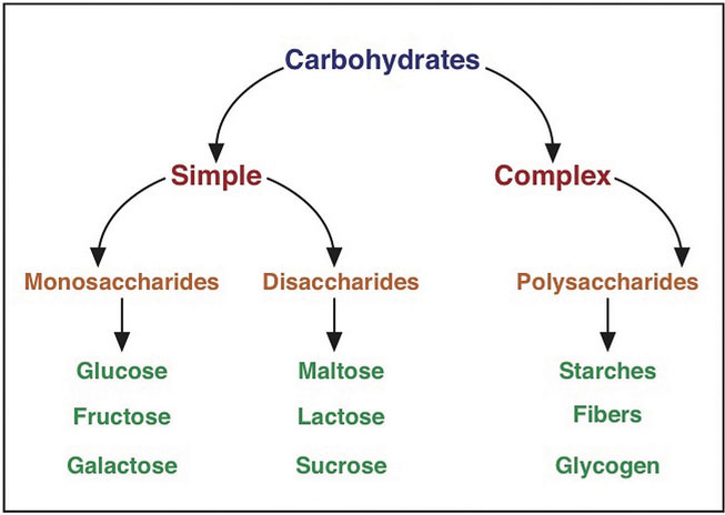
Figure 13.34 Carbohydrate Classification Scheme: Carbohydrates are broken down into the subgroups simple and complex carbohydrates. These subgroups are further categorized into mono-, di-, and polysaccharides.
Starch molecules are found in abundance in grains, legumes, and root vegetables, such as potatoes. Eating raw foods containing starches provides very little energy as the digestive system has a hard time breaking them down. Cooking breaks down the crystal structure of starches, making them much easier to break down in the human body. Starches are used widely in the food industry and during cooking as food thickeners.
Dietary fibers are polysaccharides that are highly branched and cross-linked. Some dietary fibers are pectin, gums, cellulose, hemicellulose, and lignin. Humans do not produce the enzymes that can break down dietary fiber; however, bacteria in the large intestine (colon) do. Dietary fibers are very beneficial to our health. The Dietary Guidelines Advisory Committee states that there is enough scientific evidence to support that diets high in fiber reduce the risk for obesity and diabetes, which are primary risk factors for cardiovascular disease.
Health Consequences and Benefits of High-Carbohydrate Diets