Want to create or adapt books like this? Learn more about how Pressbooks supports open publishing practices.
7 Muscular System
7.1 Introduction
In this chapter, you will learn about the muscular system, which carries out both voluntary body movements and involuntary contractions of internal organs and structures. Specifically, you will learn about:
The different types of muscle tissue — skeletal, cardiac, and smooth muscle — and their different characteristics and functions.
How muscle cells are specialized to contract and cause voluntary and involuntary movements.
The ways in which muscle contraction is controlled.
The structure and organization of skeletal muscles, including the different types of muscle fibers, and how actin and myosin filaments move across each other — according to the sliding filament theory — to cause muscle contraction.
Cardiac muscle tissue in the heart that contracts to pump blood through the body.
Smooth muscle tissue that makes up internal organs and structures, such as the digestive system, blood vessels, and uterus.
The physical and mental health benefits of aerobic and anaerobic exercise, such as running and weightlifting.
Disorders of the muscular system, including musculoskeletal disorders (such as strains and carpal tunnel syndrome) and neuromuscular disorders (such as muscular dystrophy, myasthenia gravis, and Parkinson’s disease).
7.2 INTRODUCTION TO THE MUSCULAR SYSTEM
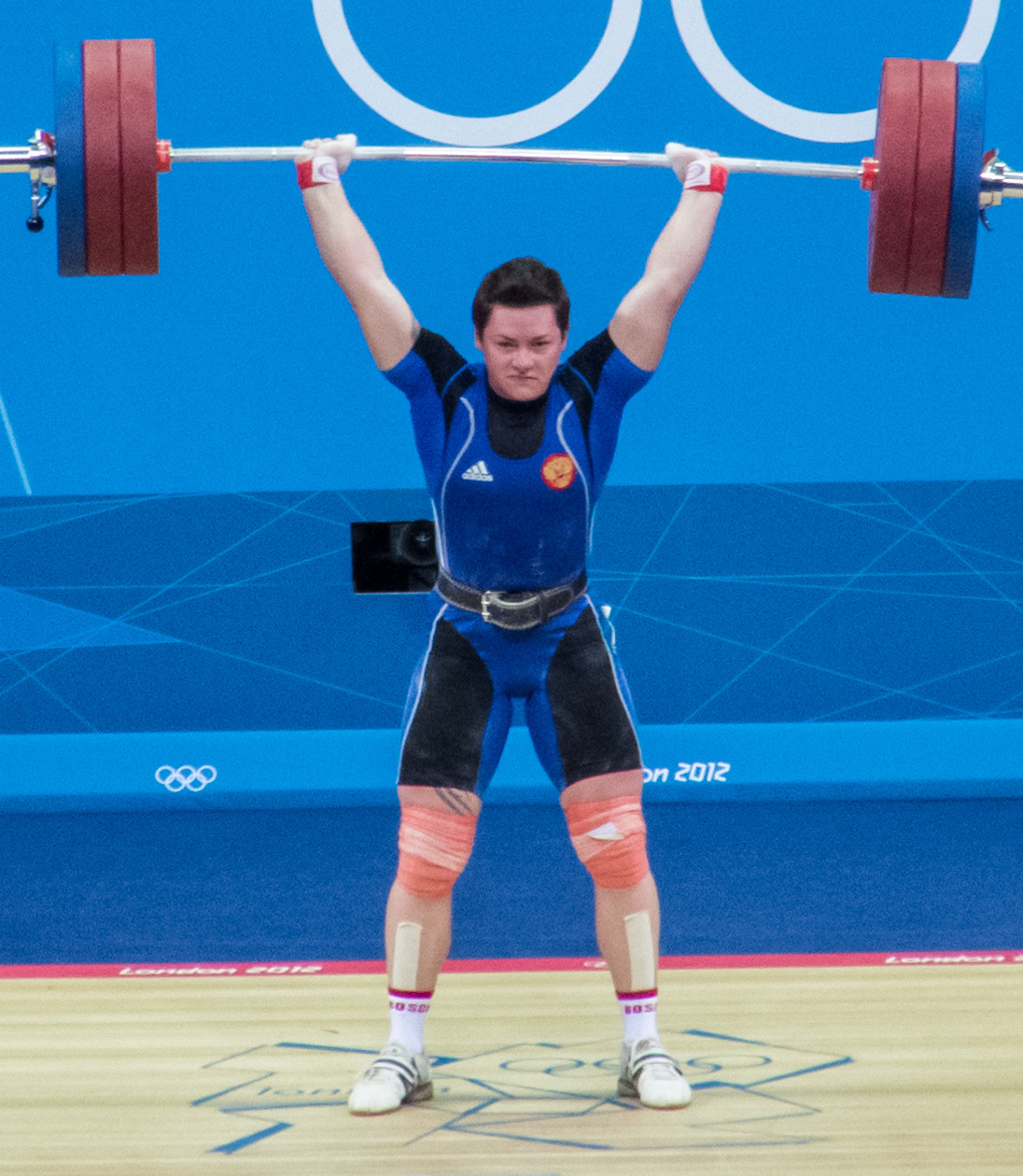
Figure 7.1 Natalia Zabolotnaya, 2012 Olympics.
MARVELOUS MUSCLES
Does the word muscle make you think of the well-developed muscles of a weightlifter, like the woman in Figure 7.1? Her name is Natalia Zabolotnaya, and she’s a Russian Olympian. The muscles that are used to lift weights are easy to feel and see, but they aren’t the only muscles in the human body. Many muscles are deep within the body, where they form the walls of internal organs and other structures. You can flex your biceps at will, but you can’t control internal muscles like these. It’s a good thing that these internal muscles work without any conscious effort on your part, because movement of these muscles is essential for survival. Muscles are the organs of the muscular system.
WHAT IS THE MUSCULAR SYSTEM?
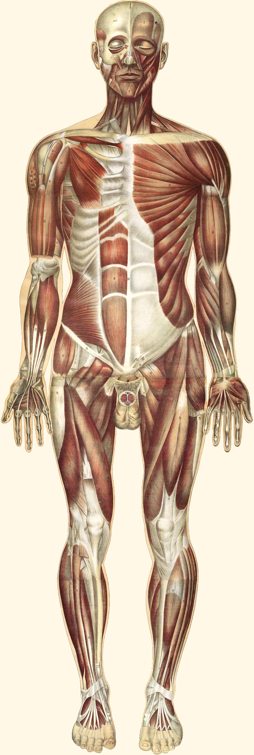
The muscular system consists of all the muscles of the body. The largest percentage of muscles in the muscular system consists of skeletal muscles, which are attached to bones and enable voluntary body movements. There are almost 650 skeletal muscles in the human body, many of them shown in Figure 7.2. Besides skeletal muscles, the muscular system also includes cardiac muscle, which makes up the walls of the heart, and smooth muscle, which control movement in other internal organs and structures.
Figure 7.2 Many of the skeletal muscles in the human muscular system are shown in this drawing of the human body.
Muscle Structure and Function
Muscles are organs composed mainly of muscle cells, which are also called muscle fibers(mainly in skeletal and cardiac muscle) or myocytes (mainly in smooth muscle). Muscle cells are long, thin cells that are specialized for the function of contracting. They contain protein filaments that slide over one another using energy in ATP. The sliding filaments increase the tension in — or shorten the length of — muscle cells, causing a contraction. Muscle contractions are responsible for virtually all the movements of the body, both inside and out.
Skeletal muscles are attached to bones of the skeleton. When these muscles contract, they move the body. They allow us to use our limbs in a variety of ways, from walking to turning cartwheels. Skeletal muscles also maintain posture and help us to keep balance.
Smooth muscles in the walls of blood vessels contract to cause vasoconstriction, which may help conserve body heat. Relaxation of these muscles causes vasodilation, which may help the body lose heat. In the organs of the digestive system, smooth muscles squeeze food through the gastrointestinal tract by contracting in sequence to form a wave of muscle contractions called peristalsis. Think of squirting toothpaste through a tube by applying pressure in sequence from the bottom of the tube to the top, and you have a good idea of how food is moved by muscles through the digestive system. Peristalsis of smooth muscles also moves urine through the urinary tract.
Cardiac muscle tissue is found only in the walls of the heart. When cardiac muscle contracts, it makes the heartbeat. The pumping action of the beating heart keeps blood flowing through the cardiovascular system.
INTERACTIONS WITH OTHER BODY SYSTEMS
Muscles cannot contract on their own. Skeletal muscles need stimulation from motor neurons in order to contract. The point where a motor neuron attaches to a muscle is called a neuromuscular junction. Let’s say you decide to raise your hand in class. Your brain sends electrical messages through motor neurons to your arm and shoulder. The motor neurons, in turn, stimulate muscle fibres in your arm and shoulder to contract, causing your arm to rise.
Involuntary contractions of smooth and cardiac muscles are also controlled by electrical impulses, but in the case of these muscles, the impulses come from the autonomic nervous system (smooth muscle) or specialized cells in the heart (cardiac muscle). Hormones and some other factors also influence involuntary contractions of cardiac and smooth muscles. For example, the fight-or-flight hormone adrenaline increases the rate at which cardiac muscle contracts, thereby speeding up the heartbeat.
Muscles cannot move the body on their own. They need the skeletal system to act upon. The two systems together are often referred to as the musculoskeletal system. Skeletal muscles are attached to the skeleton by tough connective tissues called tendons. Many skeletal muscles are attached to the ends of bones that meet at a joint. The muscles span the joint and connect the bones. When the muscles contract, they pull on the bones, causing them to move. The skeletal system provides a system of levers that allow body movement. The muscular system provides the force that moves the levers.
Review
What are the three types of muscle found in the body?
Muscle cells are also known as _____________________.
The point where a motor neuron attaches to a skeletal muscle is known as a ——————————–.
7.3 TYPES OF MUSCLE TISSUE
WHAT IS MUSCLE TISSUE?
Muscle tissue is a soft tissue that makes up most of the tissues in the muscles of the human muscular system. It has the unique ability to contract. Other tissues in muscles are connective tissues, such as tendons that attach skeletal muscles to bones and sheaths of connective tissues that cover or line muscle tissues. Only muscle tissue per se, has cells with the ability to contract.
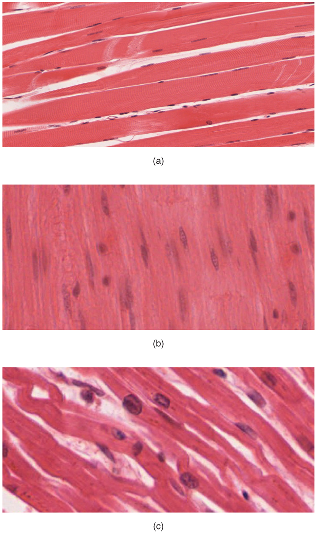
There are three major types of muscle tissues in the human body: skeletal, smooth, and cardiac muscle tissues. Figure 7.3 shows how the three types of muscle tissues appear under magnification. Below is a review of the three different tissue types.
SKELETAL MUSCLE
Skeletal muscles are voluntary muscles, meaning that you exercise conscious control over them. Skeletal muscles are attached to bones by tendons, a type of connective tissue. When these muscles shorten to pull on the bones to which they are attached, they enable the body to move. When you are exercising, reading a book, or making dinner, you are using skeletal muscles to move your body to carry out these tasks.
Under the microscope, skeletal muscles are striated (or striped) in appearance, because of their internal structure which contains alternating protein fibers of actin and myosin. Skeletal muscle is described as multinucleated, meaning one “cell” has many nuclei. This is because in utero, individual cells destined to become skeletal muscle fused, forming muscle fibers in a process known as myogenesis.
SMOOTH MUSCLE
Smooth muscles are nonstriated muscles- they still contain the muscle fibers actin and myosin, but not in the same alternating arrangement seen in skeletal muscle. Smooth muscle is found in the tubes of the body – in the walls of blood vessels and in the reproductive, gastrointestinal, and respiratory tracts. Smooth muscles are not under voluntary control meaning that they operate unconsciously, via the autonomic nervous system. Smooth muscles move substances through a wave of contraction.
CARDIAC MUSCLE
Cardiac muscles work involuntarily, meaning they are regulated by the autonomic nervous system. This is probably a good thing, since you wouldn’t want to have to consciously concentrate on keeping your heart beating all the time! Cardiac muscle, which is found only in the heart, is mononucleated and striated (due to alternating bands of myosin and actin). Their contractions cause the heart to pump blood. In order to make sure entire sections of the heart contract in unison, cardiac muscle tissue contains special cell junctions called intercalated discs, which conduct the electrical signals used to “tell” the chambers of the heart when to contract.
Figure 7.3 These magnified images show (a) skeletal muscle tissue, (b) smooth muscle tissue, and (c) cardiac muscle tissue.
Review
Where is smooth muscle found? What controls the contraction of smooth muscle?
Where is cardiac muscle found? What controls its contractions?
Where is skeletal muscle found, and what is its general function?
7.4 SKELETAL MUSCLE TISSUE
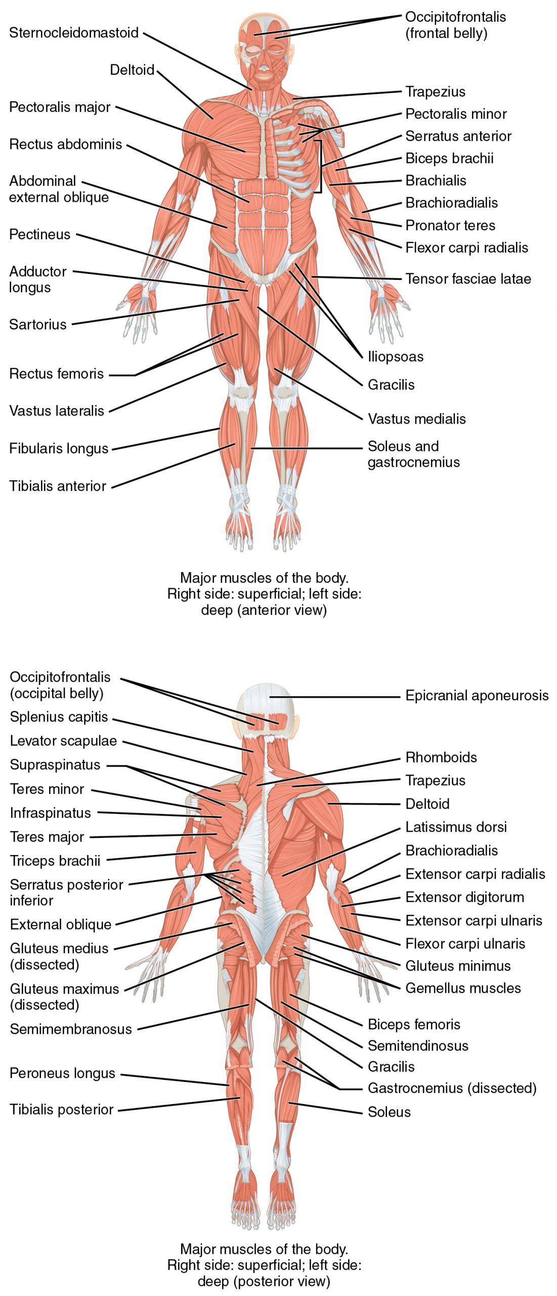
Skeletal muscle is muscle tissue that is attached to bones by tendons, which are bundles of collagen fibers. Whether you are moving your eyes or running a marathon, you are using skeletal muscles. Contractions of skeletal muscles are voluntary, or under conscious control of the central nervous system. Skeletal muscle tissue is the most common type of muscle tissue in the human body. By weight, an average adult male is about 42% skeletal muscles, and the average adult female is about 36% skeletal muscles. Some of the major skeletal muscles in the human body are labeled in Figure 7.4 below.
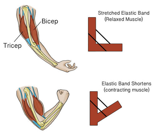
To move bones in opposite directions, skeletal muscles often consist of muscle pairs that work in opposition to one another, also called antagonistic muscle pairs. For example, when the biceps muscle (on the front of the upper arm) contracts, it can cause the elbow joint to flex or bend the arm, as shown in Figure 7.5. When the triceps muscle (on the back of the upper arm) contracts, it can cause the elbow to extend or straighten the arm. The biceps and triceps muscles, also shown in Figure 7.5, are an example of a muscle pair where the muscles work in opposition to each other. Muscle groups that work together for movement are known as synergistic muscles. An example of synergistic muscles are the rectus abdominis and the external obliques. Contraction of both allow you to bend your backbone.
Figure 7.5 Triceps and biceps muscles in the upper arm are opposing muscles that move the arm at the elbow in opposite directions.
SKELETAL MUSCLE STRUCTURE
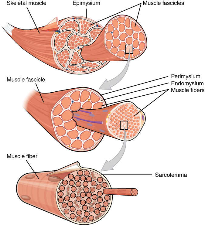
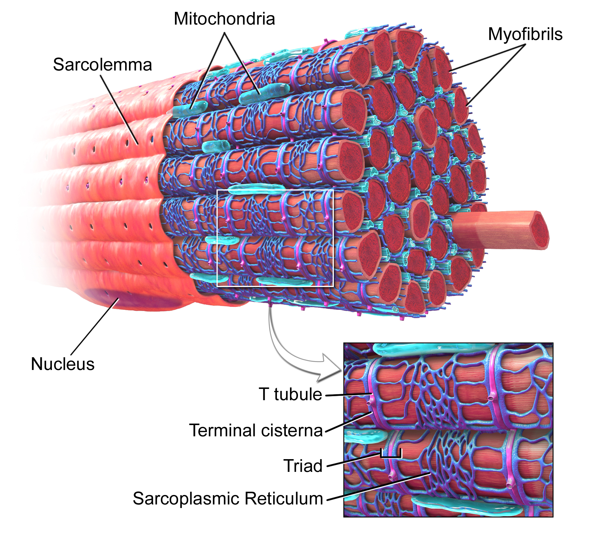
Each skeletal muscle consists of hundreds — or even thousands — of skeletal muscle fibers, which are long, string-like cells. As shown in Figure 7.6below, skeletal muscle fibers are individually wrapped in connective tissue called endomysium. The skeletal muscle fibers are bundled together in units called muscle fascicles, which are surrounded by sheaths of connective tissue called perimysium. Each fascicle contains between ten and 100 (or even more!) skeletal muscle fibers. Fascicles, in turn, are bundled together to form individual skeletal muscles, which are wrapped in connective tissue called epimysium. The connective tissues in skeletal muscles have a variety of functions. They support and protect muscle fibers, allowing them to withstand the forces of contraction by distributing the forces applied to the muscle. They also provide pathways for nerves and blood vessels to reach the muscles. In addition, the epimysium anchors the muscles to tendons.
Figure 7.6 Each skeletal muscle has a structure of bundles within bundles. Bundles of muscle fibers make up a muscle fascicle, and bundles of fascicles make up a skeletal muscle. At each level of bundling, a connective tissue membrane surrounds the bundle.
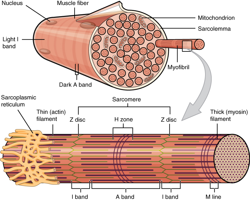
The same bundles-within-bundles structure is replicated within each muscle fiber. As shown in Figure 7.7, a muscle fiber consists of a bundle of myofibrils, which are themselves bundles of protein filaments. These protein filaments consist of thin filaments of the protein actin, which are anchored to structures called Z discs, and thick filaments of the protein myosin. The filaments are arranged together within a myofibril in repeating units called sarcomeres., which run from one Z disc to the next. The sarcomere is the basic functional unit of skeletal and cardiac muscles. It contracts as actin and myosin filaments slide over one another. Skeletal muscle tissue is said to be striated, because it appears striped. It has this appearance because of the regular, alternating A (dark) and I (light) bands of filaments arranged in sarcomeres inside the muscle fibers. Other components of a skeletal muscle fiber include multiple nuclei and mitochondria.
Figure 7.7 Bundles of protein filaments form a myofibril, and bundles of myofibrils make up a single muscle fiber. I and A bands refer to the positioning of myosin and actin fibers in a myofibril. Sarcoplasmic reticulum is a specialized type of endoplasmic reticulum that forms a network around each myofibril. It serves as a reservoir for calcium ions, which are needed for muscle contractions. H zones and Z discs are also involved in muscle contractions, which you can read about in the concept Muscle Contraction.
MUSCLE CONTRACTION
A muscle contraction is an increase in the tension or a decrease in the length of a muscle. Muscle tension is the force exerted by the muscle on a bone or other object. A muscle contraction is isometric if muscle tension changes, but muscle length remains the same. An example of isometric muscle contraction is holding a book in the same position. A muscle contraction is isotonic if muscle length changes, but muscle tension remains the same. An example of isotonic muscle contraction is raising a book by bending the arm at the elbow. The termination of a muscle contraction of either type occurs when the muscle relaxes and returns to its non-contracted tension or length.
It’s obvious that a sport like arm wrestling depends on muscle contractions. Arm wrestlers must contract muscles in their hands and arms and keep them contracted in order to resist the opposing force exerted by their opponent. The wrestler whose muscles can contract with greater force wins the match. To use our arm-wrestling example, if both arm wrestlers have equal strength and they are pulling with all their might, but there is no movement, that is isometric muscle contraction. However, as soon as one arm wrestler starts to win and is able to start pulling the opponents arm down, that is isotonic muscle contraction.
HOW A SKELETAL MUSCLE CONTRACTION BEGINS
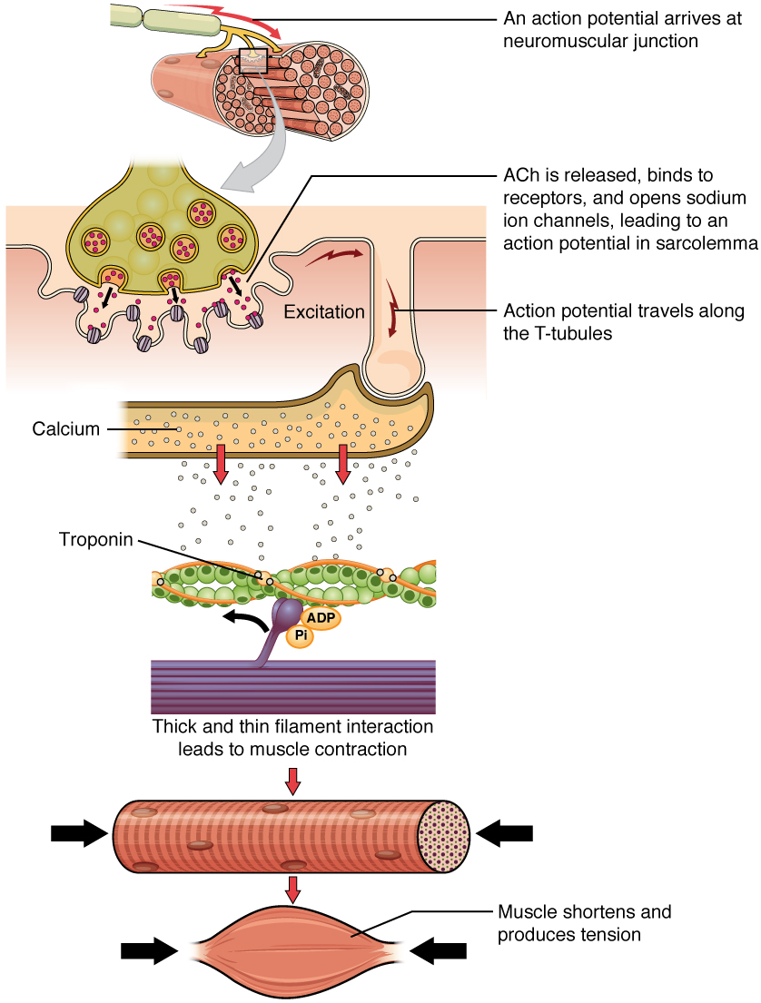
Excluding reflexes, all skeletal muscle contractions occur as a result ofconsciouseffortoriginatinginthebrain.Thebrainsends electrochemicalsignalsthroughthesomaticnervoussystemto motor neurons that innervate muscle fibers. A single motor neuron with multiple axon terminals can innervate multiple muscle fibers, thereby causing them to contract at the same time. The connection between a motor neuron axon terminal and a muscle fiber occurs at a neuromuscular junction site.This is a chemical synapse where a motor neuron transmits a signal to muscle fiber to initiate a muscle contraction.
The process by which a signal is transmitted at a neuromuscular junction is illustrated in Figure 7.8. The sequence of events begins when an action potential is initiated in the cell body of a motor neuron, and the action potential is propagated along the neuron’s axontotheneuromuscularjunction.Oncetheactionpotential reaches the end of the axon terminal, it causes the neurotransmitter acetylcholine (ACh) from synaptic vesicles in the axon terminal. The ACh molecules diffuse across the synaptic cleft and bind to the musclefiberreceptors,therebyinitiatingamusclecontraction. Musclecontractionisinitiatedwiththedepolarizationofthe sarcolemma caused by the sodium ions’ entrance through the sodium channels associated with the ACh receptors.
Figure 7.7: This diagram represents the sequence of events that occurs when a motor neuron stimulates a muscle fiber to contract. The action potential travels down the t-tubules and excites the sarcoplasmicreticulumwhichreleasescalcium.Calciumwhen bound to troponin causes conformational changes in the sarcomere. Consequently, the interaction of thick and thin filaments of the sarcomere leads to muscle contraction.
Things happen very quickly in the world of excitable membranes (think about how quickly you can snap your fingers as soon as you decidetodoit).Immediatelyfollowingdepolarizationofthe membrane, it repolarizes, andre-establishes the negative membrane potential. Meanwhile, the ACh in the synaptic cleft is degraded by the enzyme acetylcholinesterase (AChE). The ACh cannot rebind to a receptor and reopen its channel, which would cause unwanted extended muscle excitation and contraction.
Propagation of an action potential along the sarcolemma enters the T-tubules. For the action potential to reach the membrane of the Sarcoplasmic Reticulum (SR), there are periodic invaginations in the sarcolemma, calledT-tubules(“T” stands for “transverse”). The arrangement of a T-tubule with the membranes of SR on either side is called atriad(Figure 7.8). The triad surrounds the cylindrical structure called amyofibril, which contains actin and myosin. The T-tubules carry the action potential into the interior of the cell, which triggers the opening of calcium channels in the membrane of the adjacent SR, causingto calcium diffuse out of the SR and into the sarcoplasm. It is the arrival of calcium in the sarcoplasm that initiates. contraction of the muscle fiber by its contractile units, or sarcomeres.
Figure 7.8: Narrow T-tubules permit the conduction of electrical impulses. The SR functions to regulate intracellular levels of calcium. Two terminal cisternae (where enlarged SR connects to the T-tubule) and one T-tubule comprise a triad—a “threesome” of membranes, with those of SR on two sides and the T-tubule sandwiched between them.
Excitation-contraction coupling
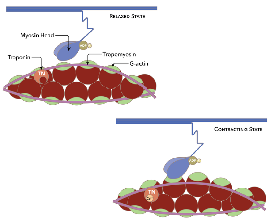
Although the termexcitation-contraction couplingconfuses or scares -some students, it comes down to this: for a skeletal muscle fiber to contract, its membrane must first be “excited”—in other words, it must be stimulated to fire an action potential. The muscle fiber action potential, which sweeps along the sarcolemma as a wave, is “coupled” to the actual contraction through the release of calcium ions (Ca++)fromtheSR.Oncereleased,the Ca++ interacts with the shielding proteins, troponin-tropomyosin complex, forcing them to move aside so that the actin-binding sites are available for attachment by myosin heads. The myosin then pulls the actin filaments toward the center, shortening the muscle fiber.
Figure 7.9: Tropomyosin Troponin complex shields the cross-bridge sites on actin. Myosin can only bind with actin when this complex is removed with the help of Calcium ions.
In skeletal muscle, this sequence begins with signals from the somatic motor division of the nervous system. In other words, the “excitation” step in skeletal muscles is always triggered by signaling from the nervous system.
SLIDING FILAMENT THEORY OF MUSCLE CONTRACTION
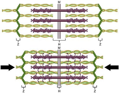
Once the muscle fiber is stimulated by the motor neuron, actin, and myosin protein filaments within the skeletal muscle fiber slide past each other to produce a contraction. Thesliding filament theoryis themostwidelyacceptedexplanationforhowthisoccurs. According to this theory, muscle contraction is a cycle of molecular events in which thick myosin filaments repeatedly attach to and pull-on thin actin filaments, so they slide over one another. The actin filaments are attached to Z discs, each of which marks the end of a sarcomere. The sliding of the filaments pulls the Z discs of a sarcomere closer together, thus shortening the sarcomere. As this occurs, the muscle contracts.
Figure 7.10: The top diagram shows a relaxed sarcomere, and the bottom diagram shows a contracted sarcomere. Please note the z discs, h zone, and M line. In a contracted sarcomere the H zone reduces as compared to relaxed sarcomere because actin fibers (greenish-yellow double helix) move towards the M line.
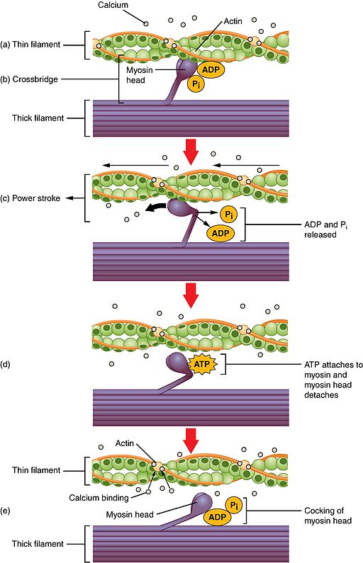
CROSSBRIDGE CYCLING
Crossbridgecyclingisasequenceofmoleculareventsthat underlies the sliding filament theory. There are many projections from the thick myosin filaments, each of which consists of two myosin heads. Each myosin head has binding sites for ATP and actin. The thin actin filaments also have binding sites for the myosin heads—a cross-bridge forms when a myosin head binds with an actin filament.
The process of cross-bridge cycling is shown in Figure 7.11. A cross-bridge cycle begins when the myosin head binds to an actin filament. ADP and Pare also bound to the myosin head at this stage. Next, a power stroke moves the actin filament inward toward the sarcomere center, thereby shortening the sarcomere. At the end of the power stroke, ADP and P are released from the myosin head, leaving the myosin head attached to the thin filament until another ATP binds to the myosin head. When ATP binds to the myosin head, it causes the myosin head to detach from the actin filament. ATP is again split into ADP and P and the energy released is used to move the myosin head into a “cocked” position. Once in this position, the myosin head can bind to the actin filament again, and another cross-bridge cycle begins.
Figure 7.11: Crossbridge cycling Interesting and hopeful basic research on muscle contraction is often in the news because muscle contractions are involved in so many different body processes and disorders, including heart failure and stroke.
Muscle Contraction 3D, 3DBiology, 2017.
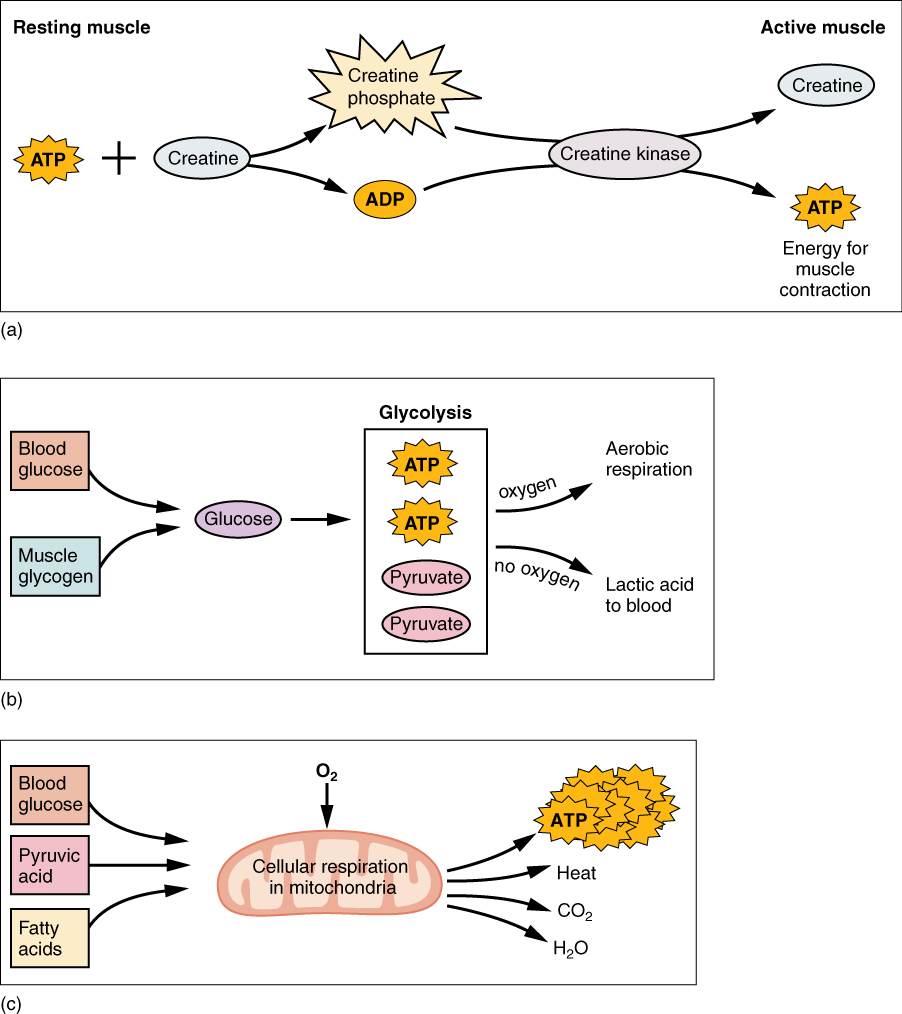
ENERGY FOR MUSCLE CONTRACTION
According to the sliding filament theory, ATP is needed to provide the energy for a muscle contraction. Where does this ATP come from? Actually, there are multiple potential sources, as illustrated in Figure 7.12 below.
As you can see from the first diagram, some ATP is already available in a resting muscle. As a muscle contraction starts, this ATP is used up in just a few seconds. More ATP is generated from creatine phosphate, but this ATP is used up rapidly as well. It’s gone in another 15 seconds or so.
Glucose from the blood and glycogen stored in muscle can then be used to make more ATP. Glycogen breaks down to form glucose, and each glucose molecule produces two molecules of ATP and two molecules of pyruvate. Pyruvate (as pyruvic acid) can be used in aerobic respiration if oxygen is available. Alternatively, pyruvate can be used in anaerobic respiration, if oxygen is not available. The latter produces lactic acid, which may contribute to muscle fatigue. Anaerobic respiration typically occurs only during strenuous exercise when so much ATP is needed that sufficient oxygen cannot be delivered to the muscle to keep up.
Resting or moderately active muscles can get most of the ATP they need for contractions by aerobic respiration. This process takes place in the mitochondria of muscle cells. In the process, glucose and oxygen react to produce carbon dioxide, water, and many molecules of ATP.
Figure 7.12 Muscles require many ATP molecules to power muscle contractions. The ATP can come from the three sources illustrated in diagrams a-c.
Interactions of Skeletal Muscles
Skeletal muscles interact to produce movements by way of anatomical positioning and the coordinated summation of innervation signals. These are the key points concerning interactions:
Muscle contractions can be termed twitch, summation or tetanus.
A twitch contraction is the period of contraction and relaxation of a muscle after a single stimulation.
Summation is the occurrence of additional twitch contractions before the previous twitch has completely relaxed.
Summation can be achieved by increasing the frequency of stimulation, or by recruiting additional muscle fibers within a muscle.
Tetanus occurs when the frequency of muscle contraction is such that the maximal force is tension is generated without any relaxation of the muscle.
Twitch
When stimulated by a single action potential a muscle contracts and then relaxes. The time between the stimulus and the initiation of contraction is termed the latent period, which is followed by the contraction period. At peak contraction the muscle relaxes and returns to its resting position. Taken all together these three periods are termed a twitch.
Figure 7.13 Muscle Twitch Contraction: The time between stimulation and contraction is termed the latent period. After contraction the muscle relaxes back to a resting level of tension. Together these three periods form a single muscle twitch,
Summation
If an additional action potential were to stimulate a muscle contraction before a previous muscle twitch had completely relaxed then it would sum onto this previous twitch increasing the total amount of tension produced in the muscle. This addition is termed summation. Within a muscle summation can occur across motor units to recruit more muscle fibers, and also within motor units by increasing the frequency of contraction.
Multiple fiber summation
When a weak signal is sent by the central nervous system to contract a muscle, the smaller motor units, being more excitable than the larger ones, are stimulated first. As the strength of the signal increases, more (and larger) motor units are excited. The largest motor units have as much as 50 times the contractile strength as the smaller ones; thus, as more and larger motor units are activated, the force of muscle contraction becomes progressively stronger. A concept known as the size principle allows for a gradation of muscle force during weak contraction to occur in small steps, which become progressively larger as greater amounts of force are required.
Frequency summation
For skeletal muscles, the force exerted by the muscle can be controlled by varying the frequency at which action potentials are sent to muscle fibers. Action potentials do not arrive at muscles synchronously, and, during a contraction, only a certain percentage of the fibers in the muscle will be contracting at any given time. In a typical circumstance, when a human is exerting as much muscular force as they are consciously able, roughly one-third of the fibers in that muscle will be contracting at once. This relatively low level of contraction is a protective mechanism to prevent damage to the muscle tissue and attaching tendons and structures.
Tetanus
If the frequency of action potentials generated increases to such a point that muscle tension has reached its peak and plateaued and no relaxation is observed, then the muscle contraction is described as a tetanus.
Figure 7.14 Summation and Tetanus Contractions: Repeated twitch contractions, where the previous twitch has not relaxed completely are called a summation. If the frequency of these contractions increases to the point where maximum tension is generated and no relaxation is observed, then the contraction is termed a tetanus.
SLOW and FAST-TWITCH SKELETAL MUSCLE FIBERS
Skeletal muscle fibers can be divided into two types, called slow-twitch (or type I) muscle fibers and fast-twitch (or type II) muscle fibers.
Slow-twitch fibers are dense with capillaries and rich in mitochondria and myoglobin, which is a protein that stores oxygen until needed for muscle activity. Relative to fast-twitch fibers, slow-twitch fibers can carry more oxygen and sustain aerobic (oxygen-using) activity. Slow-twitch fibers can contract for long periods of time, but not with very much force. They are relied upon primarily in endurance events, such as distance running or cycling.
Fast-twitch fibers contain fewer capillaries and mitochondria and less myoglobin. This type of muscle fiber can contract rapidly and powerfully, but it fatigues very quickly. Fast-twitch fibers can sustain only short, anaerobic (non-oxygen-using) bursts of activity. Relative to slow-twitch fibers, fast-twitch fibers contribute more to muscle strength and have a greater potential for increasing in mass. They are relied upon primarily in short, strenuous events, such as sprinting or weightlifting.
Proportions of fiber types vary considerably from muscle to muscle and from person to person. Individuals may be genetically predisposed to have a larger percentage of one type of muscle fiber than the other. Generally, an individual who has more slow-twitch fibers is better suited for activities requiring endurance, whereas an individual who has more fast-twitch fibers is better suited for activities requiring short bursts of power.
Review
What is muscle tissue?
Why do many skeletal muscles work in pairs?
Describe the structure of a skeletal muscle.
Relate muscle fiber structure to the functional units of muscles.
Why is skeletal muscle tissue striated?
Give one example of connective tissue that is found in muscles. Describe one of its functions.
Explain why an action potential in a single motor neuron can cause multiple muscle fibers to contract.
The name of the synapse between a motor neuron and a muscle fiber is the _______________ _________.
If a drug blocks the acetylcholine receptors on muscle fibers, what do you think this would do to muscle contraction? Explain your answer.
True or False:According to the sliding filament theory, actin filaments actively attach to and pull-on myosin filaments.
TrueorFalse:Whenamotorneuronproducesanaction potential, the sarcomeres in the muscle fiber that it innervates become shorter as a result.
What is the difference Fast-twitch and slow-twitch muscle fibers?
When does anaerobic respiration typically occur in human muscle cells?
If there were no ATP available in a muscle, how would this affect cross-bridge cycling? What would this do to
muscle contraction?
What is a muscle twitch? How would you distinguish between tetanus and summation?
7.5 PHYSICAL EXERCISE
Physical exercise is any bodily activity that enhances or maintains physical fitness and overall health and wellness. We generally think of physical exercise as activities that are undertaken for the main purpose of improving physical fitness and health. However, physical activities that are undertaken for other purposes may also count as physical exercise. Scrubbing a floor, raking a lawn, or playing active games with young children or a pet are all activities that can have fitness and health benefits, even though they generally are not done mainly for this purpose.
TYPES OF PHYSICAL EXERCISE
Physical exercise can be classified into three types, depending on the effects it has on the body: aerobic exercise, anaerobic exercise, and flexibility exercise. Many specific examples of physical exercise (including playing soccer and rock climbing) can be classified as more than one type.
AEROBIC EXERCISE
Figure 7.15 Kayaking is a form of aerobic exercise.
Aerobic exercise is any physical activity in which muscles are used at well below their maximum contraction strength, but for long periods of time. Aerobic exercise uses a relatively high percentage of slow-twitch muscle fibers that consume a large amount of oxygen. The main goal of aerobic exercise is to increase cardiovascular endurance, although it can have many other benefits, including muscle toning. Examples of aerobic exercise include cycling, swimming, brisk walking, jumping rope, rowing, hiking, tennis, and kayaking as shown in Figure 7.15.
ANAEROBIC EXERCISE
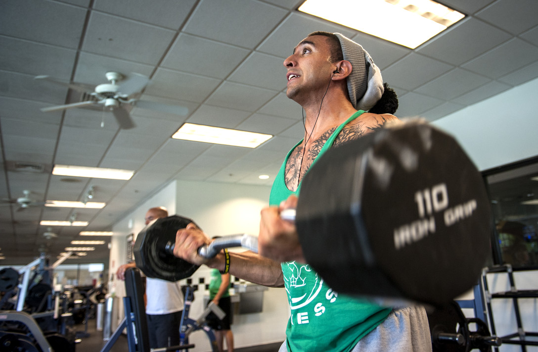
Anaerobic exercise any physical activity in which muscles are used at close to their maximum contraction strength, but for relatively short periods of time. Anaerobic exercise uses a relatively high percentage of fast-twitch muscle fibers that consume a small amount of oxygen. Goals of anaerobic exercise include building and strengthening muscles, as well as improving bone strength, balance, and coordination. Examples of anaerobic exercise include push-ups, lunges, sprinting, interval training, resistance training, and weight training (such as biceps curls with a dumbbell, as pictured in Figure 7.16).
Figure 7.16 Pitting the biceps muscle in the upper arm against a heavy weight helps to build and strengthen this muscle.
FLEXIBILITY EXERCISE
Figure 7.17 Flexibility exercise can increase range of motion and lower risk of injury.
Flexibility exercise is any physical activity that stretches and lengthens muscles. Goals of flexibility exercise include increasing joint flexibility, keeping muscles limber, and improving the range of motion, all of which can reduce the risk of injury. Examples of flexibility exercise include stretching, yoga (as in Figure 7.17), and tai chi.
Aerobic vs Anaerobic Difference, Dorian Wilson, 2017
HEALTH BENEFITS OF PHYSICAL EXERCISE
Many studies have shown that physical exercise is positively correlated with a diversity of health benefits. Some of these benefits include maintaining physical fitness, losing weight and maintaining a healthy weight, regulating digestive health, building and maintaining healthy bone density, increasing muscle strength, improving joint mobility, strengthening the immune system, boosting cognitive ability, and promoting psychological well-being. Some studies have also found a significant positive correlation between exercise and both quality of life and life expectancy. People who participate in moderate to high levels of physical activity have been shown to have lower mortality rates than people of the same ages who are not physically active and daily exercise has been shown to increase life expectancy up to an average of five years.
The underlying physiological mechanisms explaining why exercise has these positive health benefits are not completely understood. However, developing research suggests that many of the benefits of exercise may come about because of the role of skeletal muscles as endocrine organs. Contracting muscles release hormones called myokines, which promote tissue repair and the growth of new tissue. Myokines also have anti-inflammatory effects, which, in turn, reduce the risk of developing inflammatory diseases. Exercise also reduces levels of cortisol, the adrenal cortex stress hormone that may cause many health problems — both physical and mental — at sustained high levels.
CARDIOVASCULAR BENEFITS OF PHYSICAL EXERCISE
The beneficial effects of exercise on the cardiovascular system are well documented. Physical inactivity has been identified as a risk factor for the development of coronary artery disease. There is also a direct correlation between physical inactivity and cardiovascular disease mortality. Physical exercise, in contrast, has been demonstrated to reduce several risk factors for cardiovascular disease, including hypertension (high blood pressure), “bad” cholesterol (low-density lipoproteins), high total cholesterol, and excess body weight. Physical exercise has also been shown to increase “good” cholesterol (high-density lipoproteins), insulin sensitivity, the mechanical efficiency of the heart, and exercise tolerance, which is the ability to perform physical activity without undue stress and fatigue.
COGNITIVE BENEFITS OF PHYSICAL EXERCISE
Physical exercise has been shown to help protect people from developing neurodegenerative disorders, such as dementia. A 30-year study of almost 2,400 men found that those who exercised regularly had a 59 per cent reduction in dementia when compared with those who did not exercise. Similarly, a review of cognitive enrichment therapies for the elderly found that physical activity — in particular, aerobic exercise — can enhance the cognitive function of older adults. Anecdotal evidence suggests that frequent exercise may even help reverse alcohol-induced brain damage. There are several possible reasons why exercise is so beneficial for the brain. Physical exercise:
Increases blood flow and oxygen availability to the brain.
Increases growth factors that promote new brain cells and new neuronal pathways in the brain.
Increases levels of neurotransmitters (such as serotonin), which increase memory retention, information processing, and cognition.
MENTAL HEALTH BENEFITS OF PHYSICAL EXERCISE
Numerous studies suggest that regular aerobic exercise works as well as pharmaceutical antidepressants in treating mild-to-moderate depression. A possible reason for this effect is that exercise increases the biosynthesis of at least three neurochemicalsthat may act as euphoriants. The euphoric effect of exercise is well known. Distance runners may refer to it as “runner’s high,” and people who participate in crew (as in Figure 7.18) may refer to it as “rower’s high.” Because of these effects, health care providers often promote the use of aerobic exercise as a treatment for depression.
Figure 7.18 These rowing duos are competing in the 2016 Summer Olympics in Rio, in which Canada won a silver medal. They are clearly exerting themselves — and no doubt increasing their euphoriant neurochemicals in the process.
Additional mental health benefits of physical exercise include reducing stress, improving body image, and promoting positive self-esteem. Conversely, there is evidence to suggest that being sedentary is associated with increased risk of anxiety.
SLEEP BENEFITS OF PHYSICAL EXERCISE
A recent review of published scientific research suggests that exercise generally improves sleep for most people, and helps sleep disorders, such as insomnia. In fact, exercise is the most recommended alternative to sleeping pills for people with insomnia. For sleep benefits, the optimum time to exercise may be four to eight hours before bedtime, although exercise at any time of day seems to be beneficial. The only possible exception is heavy exercise undertaken shortly before bedtime, which may actually interfere with sleep.
OTHER BENEFITS OF PHYSICAL EXERCISE
Some studies suggest that physical activity may benefit the immune system. For example, moderate exercise has been found to be associated with a decreased incidence of upper respiratory tract infections. Evidence from many studies has found a correlation between physical exercise and reduced death rates from cancer, specifically breast cancer and colon cancer. Physical exercise has also been shown to reduce the risk of type 2 diabetes and obesity.
VARIATION IN RESPONSES TO PHYSICAL EXERCISE
Figure 7.19 This participant in the Toronto Marathon is likely to have a relatively high proportion of slow-twitch muscle fibres that increase her endurance.
Not everyone benefits equally from physical exercise. When participating in aerobic exercise, most people will have a moderate increase in their endurance, but some people will as much as double their endurance. Some people, on the other hand, will show little or no increase in endurance from aerobic exercise. Genetic differences in slow-twitch and fast-twitch skeletal muscle fibers may play a role in these different results. People with more slow-twitch fibers may be able to develop greater endurance, because these muscle fibers have more capillaries, mitochondria and myoglobin than fast-twitch fibers. As a result, slow-twitch fibers can carry more oxygen and sustain aerobic activity for a longer period of time than fast-twitch fibers. Studies show that endurance athletes (like the marathoner pictured in Figure 7.19) generally do tend to have a higher proportion of slow-twitch fibers than other people.
There is also great variation in individual responses to muscle building as a result of anaerobic exercise. Some people have a much greater capacity to increase muscle size and strength, whereas other people never develop large muscles, no matter how much they exercise them. People who have more fast-twitch than slow-twitch muscle fibers may be able to develop bigger, stronger muscles, because fast-twitch muscle fibers contribute more to muscle strength and have greater potential to increase in mass. Evidence suggests that athletes who excel at power activities (such as throwing and jumping) tend to have a higher proportion of fast-twitch fibers than do endurance athletes.
CAN YOU “OVERDOSE” ON PHYSICAL EXERCISE?
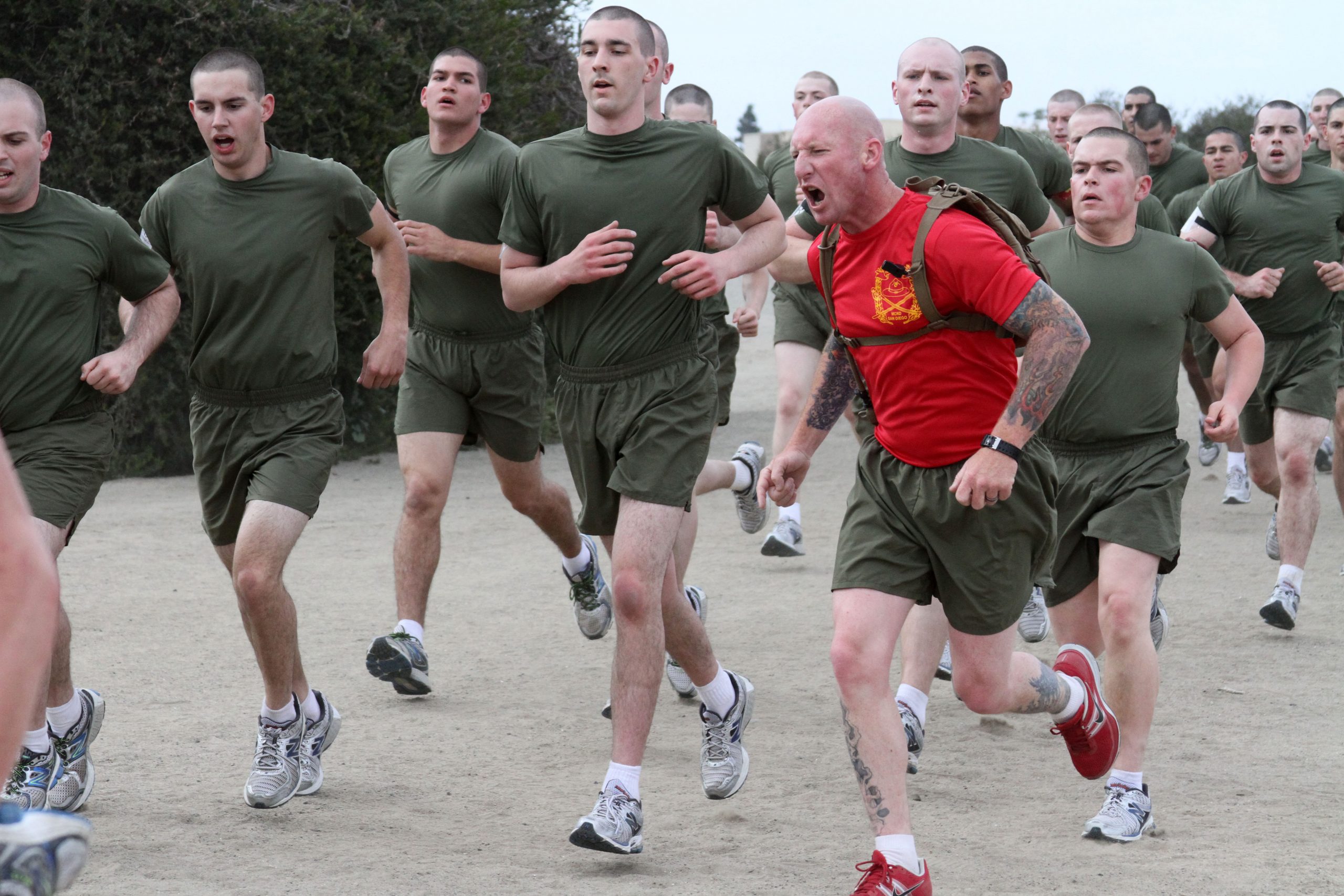
Is it possible to exercise too much? Can too much exercise be harmful? Evidence suggests that some adverse effects may occur if exercise is extremely intense and the body is not given proper rest between exercise sessions. Athletes who train for multiple marathons have been shown to develop scarring of the heart and heart rhythm abnormalities. Doing too much exercise without prior conditioning also increases the risk of injuries to muscles and joints. Damage to muscles due to overexertion is often seen in new military recruits (see Figure 7.20). Too much exercise in females may cause amenorrhea, which is a cessation of menstrual periods. When this occurs, it generally indicates that a woman is pushing her body too hard.
Figure 7.20 New military recruits may suffer muscle damage from overexertion of unconditioned muscles. The drill instructor pictured here (in orange shirt) is doing his best to encourage these marine recruits to expend their maximum effort.
Many people develop delayed onset muscle soreness (DOMS), which is pain or discomfort in muscles that is felt one to three days after exercising, and generally subsides two or three days later. DOMS was once thought to be caused by the buildup of lactic acid in the muscles. Lactic acid is a product of anaerobic respiration in muscle tissues. However, lactic acid disperses fairly rapidly, so it is unlikely to explain pain experienced several days after exercise. The current theory is that DOMS is caused by tiny tears in muscle fibers, which occur when muscles are used at too high a level of intensity.
FEATURE: MY HUMAN BODY
Most people know that exercise is important for good health, and it’s easy to find endless advice about exercise programs and fitness plans. What is not so easy to find is the motivation to start exercising — and to stick with it. This is the main reason why so many people fail to get regular exercise. Practical concerns like a busy schedule and bad weather can certainly make exercising more of a challenge, but the biggest barriers to adopting a regular exercise routine are mental. If you want to exercise but find yourself making excuses or getting discouraged and giving up, here are some tips that may help you get started and stay moving:
Avoid an all-or-nothing point of view. Don’t think you need to spend hours sweating at the gym or training for a marathon to get healthy. Even a little bit of exercise is better than nothing at all. Start out with ten or 15 minutes of moderate activity each day. Taking a walk around your neighborhood is a great way to begin! From there, gradually increase the amount of time until you are exercising to at least 30 minutes a day, five days a week. Make it a routine.
Be kind to yourself, and reinforce positive behaviors with rewards. Don’t be down on yourself because you are overweight or out of shape. Don’t beat yourself up because of a supposed lack of willpower. Instead, look at any past failures as opportunities to learn and do better. When you do achieve even small exercise goals, treat yourself to something special. Did you just complete your first workout? Reward yourself with a relaxing bath or other treat.
Don’t make excuses for not exercising. Common complaints include being too busy or tired or not athletic enough. Such excuses are not valid reasons to avoid exercising, and they will sabotage any plans to improve your fitness. If you can’t find a 30-minute period to work out, try to find ten minutes, three times a day. If you’re feeling tired, know that exercise can actually reduce fatigue and boost your energy level. If you feel clumsy and uncoordinated, remind yourself that you don’t need to be athletic to take a walk or engage in vigorous house or yard work.
Find an activity that you truly enjoy doing. Don’t think you have to lift weights or run on a treadmill to exercise your muscles. If you find such activities boring or unpleasant, you won’t stick with them. Any activity that increases your heart rate and uses large muscles can provide a workout, especially if you’re not in the habit of exercising, so find something you like to do. Do you like to dance? Put on some music and dance up a sweat! Do you enjoy gardening? Get out in the yard and dig up some dirt! Still not interested? Try an activity-based video game, such as Wii or Kinect. You may find it so much fun that it doesn’t seem like exercise until you realize you’ve worked up a sweat.
Make yourself accountable. Tell friends and family members that you’re going to start exercising. You’ll be letting them — as well as yourself — down if you don’t follow through. Some people find that keeping an exercise log to track their progress is a good way to be accountable and stick to an exercise program. Perhaps the best way to keep at it is to find an exercise partner. If you’ve got someone waiting to exercise with you, you will be less likely to make excuses for not exercising.
Add more physical activity to your daily life. You don’t need to follow a structured exercise program to increase your activity level. Do your house or yard work briskly for a workout. Park your car further than necessary from work or the mall, and walk the extra distance. If you live close enough, leave the car at home and walk to and from your destination. Rather than taking elevators or escalators, walk up and down stairs. When you take breaks at work, take a walk instead of sitting. Every time a commercial comes on while you’re watching TV, take a quick exercise break — run in place or do some curls with hand weights.
Review
How do we define physical exercise?
What are current recommendations for physical exercise for adults?
Define flexibility exercise and state its benefits. What are two examples of flexibility exercises?
In general, how does physical exercise affect health, quality of life, and longevity?
What mechanism may underlie many of the general health benefits of physical exercise?
Relate physical exercise to cardiovascular disease risk.
What may explain the positive benefits of physical exercise on cognition?
How does physical exercise compare with antidepressant drugs in the treatment of depression?
Identify several other health benefits of physical exercise.
Explain how genetics may influence the way individuals respond to physical exercise.
Can too much physical exercise be harmful?
The surprising reason our muscles get tired – Christian Moro, TED-Ed, 2019.
7.6 SMOOTH MUSCLE
Smooth muscle is muscle tissue in the walls of internal organs and other internal structures such as blood vessels. When smooth muscles contract, they help the organs and vessels carry out their functions. When smooth muscles in the stomach wall contract, for example, they squeeze the food inside the stomach, helping to mix and churn the food and break it into smaller pieces. This is an important part of digestion. Contractions of smooth muscles are involuntary, so they are not under conscious control. Instead, they are controlled by the autonomic nervous system, hormones, neurotransmitters, and other physiological factors.
STRUCTURE OF SMOOTH MUSCLE
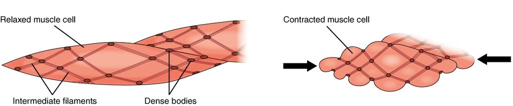
The cells that make up smooth muscle are generally called myocytes.Unlike the muscle fibers of striated muscle tissue, the myocytes of smooth muscle tissue do not have their filaments arranged in sarcomeres. Therefore, smooth tissue is not striated. However, the myocytes of smooth muscle do contain myofibrils, which in turn contain bundles of myosin and actin filaments. The filaments cause contractions when they slide over each other, as shown in Figure 7.21.
Figure 7.21The basic mechanism of muscle contraction in smooth muscle is the same as that in other types of muscle tissue.
Functions of Smooth Muscle
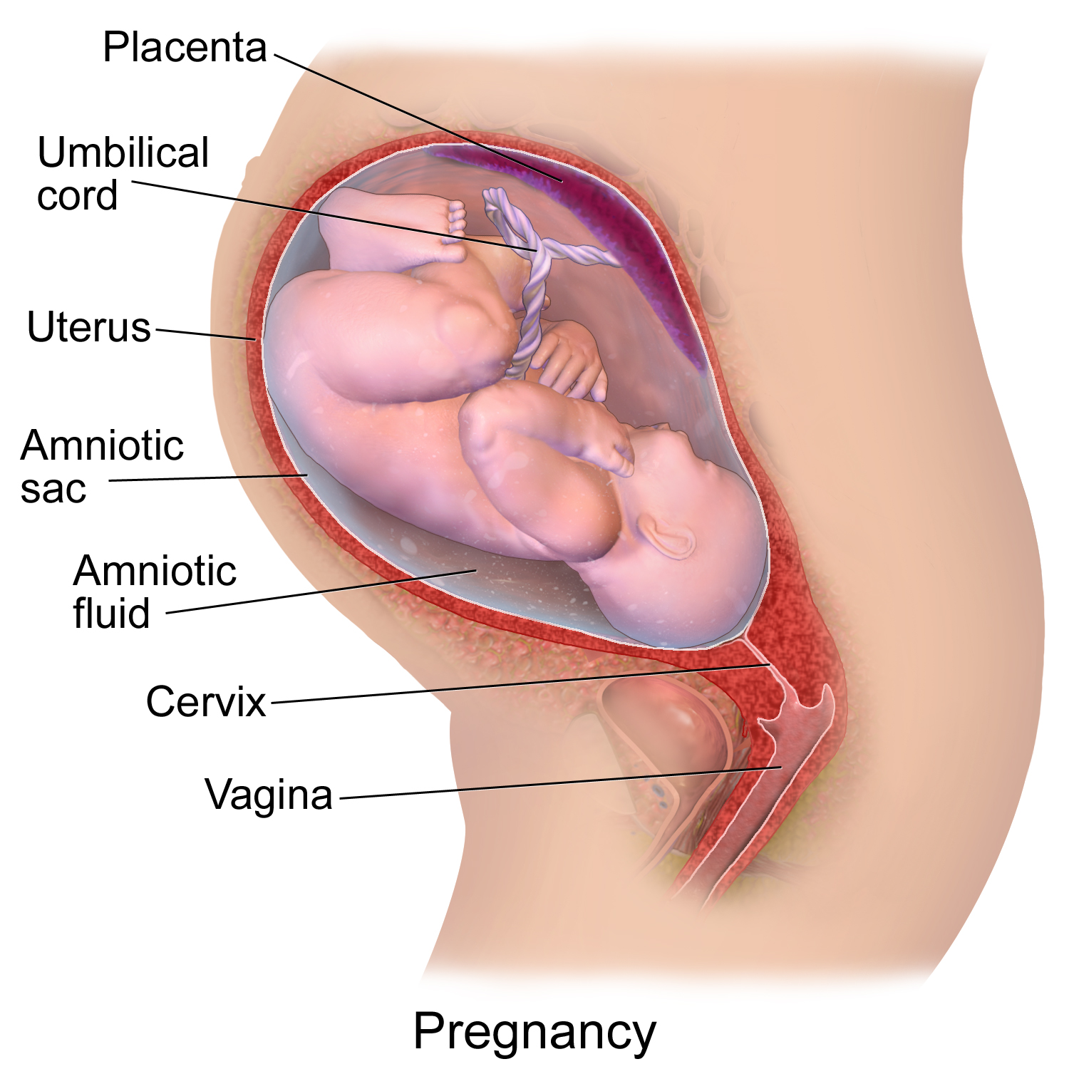
Unlike striated muscle, smooth muscle can sustain very long-term contractions. Smooth muscle can also stretch and still maintain its contractile function, which striated muscle cannot. The elasticity of smooth muscle is enhanced by an extracellular matrix secreted by myocytes. The matrix consists of elastin and collagen, and other stretchy fibers. The ability to stretch and still contract is an important attribute of smooth muscle in organs such as the stomach and uterus (see Figure 7.22), both of which must stretch considerably as they perform their normal functions.
Figure 7.22 The muscular uterine wall stretches to a great extent to accommodate a growing fetus, yet it can still contract with great force during the labor that precedes childbirth. At that time, it can exert up to 100 pounds of force.
The following list indicates where many smooth muscles are found, along with some of their specific functions.
Walls of organs of the gastrointestinal tract (such as the esophagus, stomach, and intestines), moving food through the tract by peristalsis
Walls of air passages of the respiratory tract (such as the bronchi), controlling the diameter of the passages and the volume of air that can pass through them
Walls of organs of the male and female reproductive tracts; in the uterus, for example, pushing a baby out of the uterus and into the birth canal
Walls of structures of the urinary system, including the urinary bladder, allowing the bladder to expand so it can hold more urine, and then contract as urine is released
Walls of blood vessels, controlling the diameter of the vessels and thereby affecting blood flow and blood pressure
Walls of lymphatic vessels, squeezing the fluid called lymph through the vessels
Iris of the eyes, controlling the size of the pupils and thereby the amount of light entering the eyes
Arrector pili in the skin, raising hairs in hair follicles in the dermis
7.7 CARDIAC MUSCLE
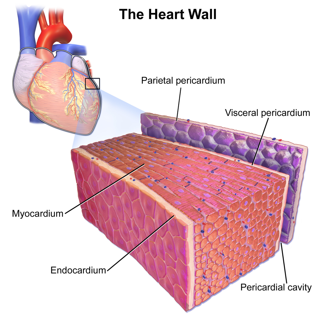
Figure 7.23 The thick wall of the heart consists mainly of cardiac muscle tissue called myocardium.
Cardiac muscle is found only in the wall of the heart. It is also called myocardium. As shown in Figure 7.23, myocardium is enclosed within connective tissues, including the endocardium on the inside of the heart and pericardium on the outside of the heart. When cardiac muscle contracts, the heart beats and pumps blood. Contractions of cardiac muscle are involuntary, like those of smooth muscles. They are controlled by electrical impulses from specialized cardiac muscle cells in an area of the heart muscle called the sinoatrial node.
Like skeletal muscle, cardiac muscle is striated because its filaments are arranged in sarcomeres inside the muscle fibers. However, in cardiac muscle, the myofibrils are branched at irregular angles rather than arranged in parallel rows (as they are in skeletal muscle). This explains why cardiac and skeletal muscle tissues look different from one another.
The cells of cardiac muscle tissue are arranged in interconnected networks. This arrangement allows rapid transmission of electrical impulses, which stimulate virtually simultaneous contractions of the cells. This enables the cells to coordinate contractions of the heart muscle.
The heart is the muscle that performs the greatest amount of physical work in the course of a lifetime. Although the power output of the heart is much less than the maximum power output of some other muscles in the human body, the heart does its work continuously over an entire lifetime without rest. Cardiac muscle contains a great many mitochondria, which produce ATP for energy and help the heart resist fatigue.
7.8 DISORDERS OF THE MUSCULAR SYSTEM
Figure 7.24 Devices can be a pain in the neck – literally.
Pain in the Neck
Spending hours each day looking down at hand-held devices is a pain in the neck — literally. The weight of the head bending forward can put a lot of strain on neck muscles, and muscle injuries can be very painful. Neck pain is one of the most common of all complaints that bring people to the doctor’s office. In any given year, about one in five adults will suffer from neck pain. That’s a lot of pains in the neck! Not all of them are due to muscular disorders, but many of them are. Muscular disorders, in turn, generally fall into two general categories: musculoskeletal disorders and neuromuscular disorders.
MUSCULOSKELETAL DISORDERS
Musculoskeletal disorders are injuries that occur in muscles or associated tissues (such as tendons) because of biomechanical stresses. They may be caused by sudden exertion, over-exertion, repetitive motions, or long periods of maintaining awkward positions. Musculoskeletal disorders are often work- or sports-related, and generally just one or a few muscles are affected. They can often be treated successfully, and full recovery can be very likely. The disorders include muscle strains, tendinitis, and carpal tunnel syndrome.
MUSCLE STRAIN
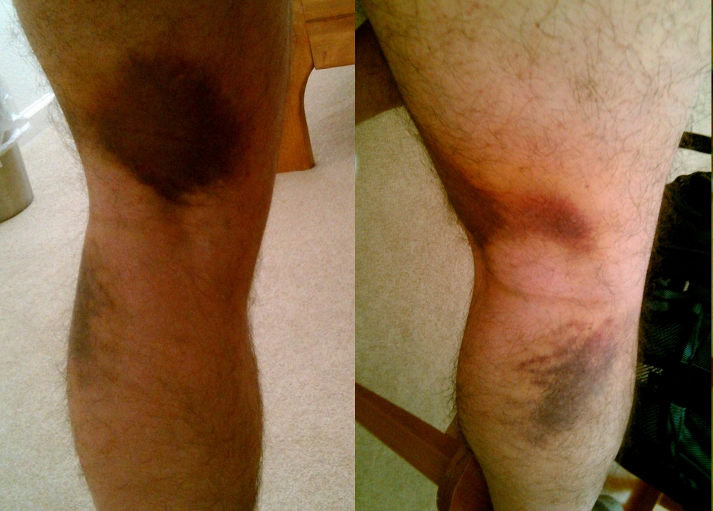
Figure 7.25 Bleeding in muscle tissue may cause a muscle strain to produce a bruise over the affected muscle, as in this hamstring bruise. The photos show two images of the same leg, both taken four days after the injury occurred.
A muscle strain is an injury in which muscle fibers tear as a result of overstretching. A muscle strain is also commonly called a pulled muscle or torn muscle. (Strains are often confused with sprains, which are similar injuries to ligaments.) Depending on the degree of injury to muscle fibers, a muscle strain can range from mildly to extremely painful. Besides pain, typical symptoms include stiffness and bruising in the area of the strained muscle. The photo here shows a large bruise caused by a hamstring muscle strain. Hamstring strains are very common in track and field athletes. In sprinters, for example, about one third of injuries are hamstring injuries. Having a previous hamstring injury puts an athlete at increased risk for having another one.
Proper first aid for a muscle strain includes five steps, which are represented by the acronym PRICE. The PRICE steps should be followed for several days after the injury. The five steps are:
Protection: Apply soft padding to the strained muscle to minimize impact with objects that might cause further damage.
Rest: Rest the muscle to accelerate healing and reduce the potential for re-injury.
Ice: Apply ice for 20 minutes at a time every two hours to reduce swelling and pain.
Compression: Apply a stretchy bandage to the strained muscle to reduce swelling.
Elevation: Keep the strained muscle elevated to reduce the chance of blood pooling in the muscle.
Non-steroidal anti-inflammatory drugs (NSAIDs, such as ibuprofen) can help reduce inflammation and relieve pain. Because such drugs interfere with blood clotting, however, they should be taken only after bleeding in the muscle has stopped — not immediately after the injury occurs. For severe muscle strains, professional medical care may be needed.
TENDINITIS
Tendinitis is inflammation of a tendon that occurs when it is over-extended or worked too hard without rest. Tendons that are commonly affected include those in the ankle, knee, shoulder, and elbow. The affected tendon depends on the type of use that causes the inflammation. Rock climbers tend to develop tendinitis in their fingers, while basketball players are more likely to develop tendinitis in the knees, to name a few examples.
Symptoms of tendinitis may include aching, sharp pain, a burning sensation, or joint stiffness. In some cases, swelling occurs around the inflamed tendon, and the area feels hot and looks red. Treatment includes the PRICE guidelines listed above, as well as the use of NSAIDs to further reduce inflammation and pain. Although symptoms should show improvement within a few days of treatment, full recovery may take several months. A gradual return to exercise or other use of the affected tendon is recommended. Physical or occupational therapy may speed the return to normal activity levels.
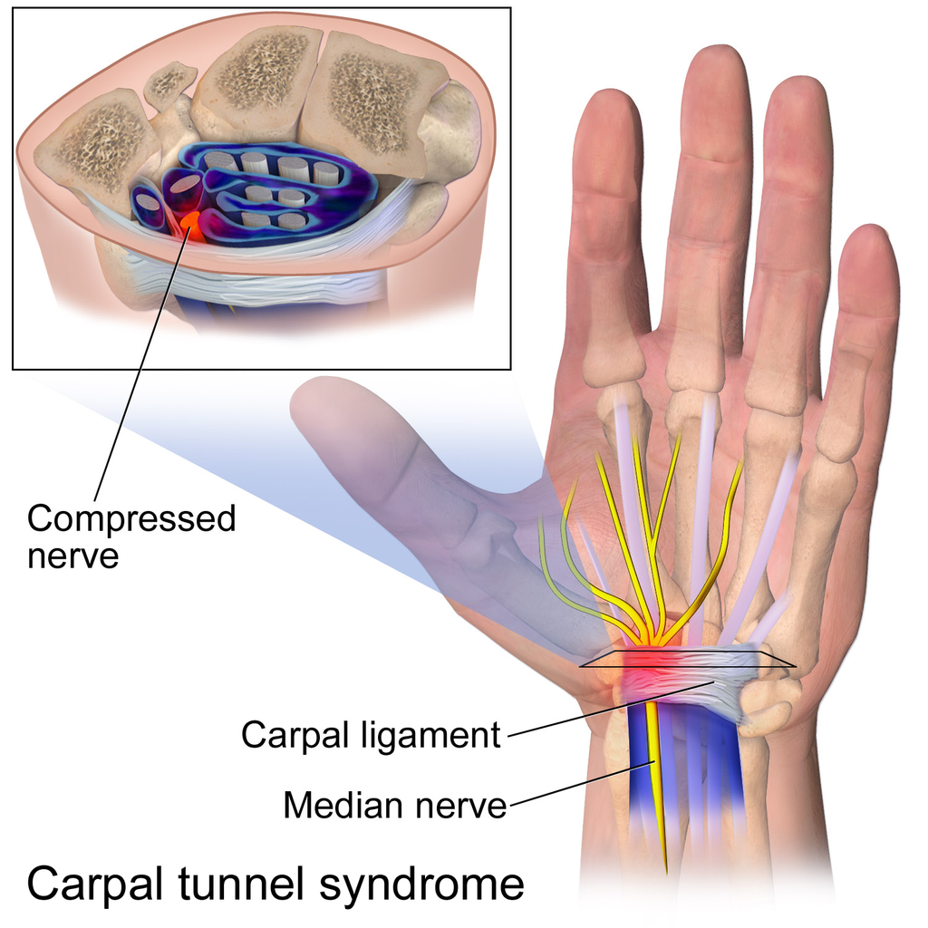
CARPAL TUNNEL SYNDROME
Carpal Tunnel Syndrome is a common biomechanical problem that occurs in the wrist when the median nerve becomes compressed between carpal bones (see Figure 7.26). This may occur due to repetitive use of the wrist, a tumor, or trauma to the wrist. Two-thirds of cases are work-related. Computer work, work with vibrating tools, and work that requires a strong grip all increase one’s risk of developing this problem. Carpal tunnel syndrome occurs more often in women than men. Other risk factors include obesity, pregnancy, and arthritis. Genetics may also play a role.
Figure 7.26 Carpal tunnel syndrome occurs when the median nerve in the wrist becomes compressed.
Compression of the median nerve results in inadequate nervous stimulation of the muscles in the thumb and first two fingers of the hand. The main symptoms are pain, numbness, and tingling in these digits. Sometimes, symptoms can be improved by wearing a wrist splint or receiving corticosteroid injections. Surgery to cut the carpal ligament reduces pressure on the median nerve and is generally more effective than nonsurgical treatment. Recurrence of carpal tunnel syndrome after surgery is rare. Without treatment, on the other hand, the lack of nervous stimulation by the median nerve may eventually cause the affected muscles of the hand to weaken and waste away.
NEUROMUSCULAR DISORDERS
Neuromuscular disorders are systemic disorders that occur because of problems with the nervous control of muscle contractions, or with the muscle cells themselves. These disorders are often due to faulty genes and not due to biomechanical stresses. Other system-wide problems, such as abnormal immune system responses, may also be involved in neuromuscular disorders.
Unlike musculoskeletal disorders, neuromuscular disorders generally affect most or all of the muscles in the body. The disorders also tend to be progressive and incurable. However, in most cases, treatment is available to slow the disease progression or to lessen the symptoms. Examples of neuromuscular disorders include muscular dystrophy, myasthenia gravis, and Parkinson’s disease.
MUSCULAR DYSTROPHY
Muscular dystrophy is a genetic disorder caused by defective proteins in muscle cells. It is characterized by progressive skeletal muscle weakness and death of muscle cells and tissues. Muscles become increasingly unable to contract in response to nervous stimulation.
There are at least nine major types of muscular dystrophy caused by different gene mutations. The most common type of childhood muscular dystrophy is Duchenne muscular dystrophy, which is due to a mutation in a recessive gene on the X chromosome. As an X-linked recessive disorder, Duchenne muscular dystrophy occurs almost exclusively in males.
Different types of muscular dystrophy affect different major muscle groups. In Duchenne muscular dystrophy, the lower limbs are affected. Signs of the disorder usually become apparent when a child starts walking. Difficulty walking becomes progressively worse through childhood. By the time a child is ten, braces may be needed for walking — and walking may no longer even by possible by age 12. The lifespan of someone with muscular dystrophy is likely to be shorter than normal because of the disease, ranging from 15 to 45 years.
In some cases, physical therapy, drug therapy, or orthopedic surgery may improve some of the signs and symptoms of muscular dystrophy. However, at present, there is no known cure for the disorder.
MYASTHENIA GRAVIS
Myasthenia gravis is a genetic neuromuscular disorder characterized by fluctuating muscle weakness and fatigue. It occurs more commonly in women, and generally begins between the ages of 20 and 40. The initial symptom of myasthenia gravis is painless muscle weakness, generally in muscles around the eye (see photos in Figure 7.27). The disease then progresses to muscles elsewhere in the body, eventually involving most of the muscles. Swallowing and chewing may become difficult as the disease progresses, and speech may become slow and slurred. In more advanced cases, myasthenia crises may occur, during which the muscles that control breathing may be affected. Emergency medical care to provide assisted ventilation is required to sustain life. A myasthenia gravis crisis may be triggered by various stressors, such as infection, fever, or stress.
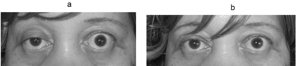
Figure 7.27 The photograph on the left shows a myasthenia gravis patient with a drooping eye lid, one of the most common signs of the disease because of weakening of muscles around the eye. The photo on the right shows the same patient after administration of a drug that blocks the breakdown of acetylcholine.
Most commonly, myasthenia gravis is caused by immune system antibodies blocking acetylcholine receptors on muscle cells, as well as the actual loss of acetylcholine receptors. Acetylcholine is the main neurotransmitter used by motor neurons to carry their signals to the muscle fibers they control. With acetylcholine blocked or the receptors lost, muscle cells fail to receive nervous stimulation to contract. Treatment of myasthenia gravis may include medications to counter the effects of the mutant gene or to suppress the immune system.
PARKINSON’S DISEASE
Parkinson’s disease is a degenerative disorder of the central nervous system that mainly affects the muscular system and movement. Four motor signs and symptoms are considered defining in Parkinson’s disease: muscle tremor (shaking), muscle rigidity, slowness of movement, and postural instability. Tremor is the most common and obvious symptom, and it most often occurs in a limb that is at rest, so it disappears during sleep or when the patient moves the limb voluntarily. Difficulty walking eventually develops, and dementia is common in the advanced stages of the disease. Depression is common, as well.
See the video “Neurology – Topic 14 – Parkinsons disease – examining a patient” by UCD Medicine, of a physician examining a patient living with advanced Parkinson’s disease:
Parkinson’s disease is more common in older people, with most cases being diagnosed after the age of 50. Often, the disease occurs for no known reason. Cases like this are called primary Parkinson’s disease. Sometimes, the disease has a known or suspected cause, such as exposure to toxins in pesticides, or repeated head trauma. In this case, it is called secondary Parkinson’s disease.
Regardless of the cause, the motor symptoms of the disease result from the death of neurons in the midbrain. The cause of cell death is not fully understood, but it appears to involve the buildup in the brain of protein structures called Lewy bodies. Early in the course of the illness, medications can be prescribed to help reduce the motor disturbances. As the disease progresses, however, the medications become ineffective. They also cause a negative side effect of involuntary writhing movements.
FEATURE: HUMAN BIOLOGY IN THE NEWS
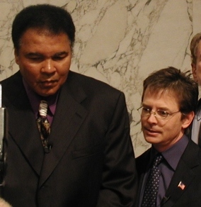
On June 3, 2016, media all over the world exploded with news of the death of Muhammad Ali at the age of 74. The world champion boxer and Olympic gold medalist died that day of complications of a respiratory infection, but the underlying cause was Parkinson’s disease. Ali was diagnosed with Parkinson’s in 1984 when he was only 42 years old. Doctors attributed his disease to repeated head trauma from boxing.
In the days following Ali’s death, the news was full of stories and images from milestones in the athlete’s life, both before and after his diagnosis with Parkinson’s disease. Sadly, the news coverage also provided an overview of his gradual decline as the disease progressed. Ali was pictured in 1996 lighting the flame at the Summer Olympics in Atlanta; however, in 2012, Ali had to be helped to his feet by his wife just to stand before the flag he was supposed to carry into the stadium. He was unable to carry it because of the ravages of Parkinson’s disease.
Figure 7.28 Muhammad Ali (left) and Michael J. Fox (right) became unlikely allies in the fight against Parkinson’s Disease, testifying together before a Senate Committee in 2002.
Muhammad Ali retired from boxing in 1981 at the age of 39, but he didn’t retire from fighting. Up until the final year of his life, Ali was a passionate activist for peace and justice, and against war and racism. In 1998, he joined Michael J. Fox, who also has Parkinson’s disease, to raise awareness of and funding for research on Parkinson’s disease. In 2002, Fox and Ali made a joint appearance before Congress to present their case. In 2005, Ali received the Presidential Medal of Freedom for the many achievements and contributions he made throughout his amazing life, in spite of Parkinson’s disease.
Review
What are musculoskeletal disorders? What causes them?
How does a muscle strain occur?
Define tendinitis. Why does it occur?
Identify first-aid steps for treating musculoskeletal disorders, such as muscle strains and tendinitis.
Describe carpal tunnel syndrome and how it may be treated.
Define neuromuscular disorders.
Identify the cause and symptoms of muscular dystrophy.
Outline the cause and progression of myasthenia gravis.
What is Parkinson’s disease? List four characteristic signs of the disorder.
What are the main differences between musculoskeletal disorders and neuromuscular disorders?
Why is padding of a strained muscle part of the typical treatment?
What are two tissues — other than muscle tissue — that can experience problems that result in muscular system disorders?
Why sitting is bad for you – Murat Dalkilinç, TED-Ed, 2015.
7.9 CASE STUDY: MUSCLES AND MOVEMENT
Figure 7.29 Who’s a good boy?
CASE STUDY: NEEDING TO RELAX
This dog (Figure 7.29) is expressing his interest in something — perhaps a piece of food — by using the neck muscles to tilt its head in an adorable fashion. Humans also sometimes tilt their heads to express interest. But imagine how disturbing and painful it would be if your neck tilted involuntarily, without you being able to control it! Forty-three-year-old Edward unfortunately knows just how debilitating this can be.
Edward has a rare condition called cervical dystonia, which is also called spasmodic torticollis. In this condition, the muscles in the neck contract involuntarily, often causing the person’s head to twist to one side. Figure 7.30 shows one type of abnormal head positioning that can be caused by cervical dystonia. The muscles may contract in a sustained fashion, holding the head and neck in one position, or they may spasm repeatedly, causing jerky movements of the head and neck.
Figure 7.30 Dystonia is a movement disorder in which a person’s muscles contract uncontrollably. The contraction causes the affected body part to twist involuntarily, resulting in repetitive movements or abnormal postures. Dystonia can affect one muscle, a muscle group, or the entire body.
Cervical dystonia is painful and can significantly interfere with a person’s ability to carry out their usual daily activities. In Edward’s case, he can no longer drive a car, because his uncontrollable head and neck movements and abnormal head positioning prevent him from navigating the road safely. He also has severe neck and shoulder pain much of the time.
Although it can be caused by an injury, there is no known cause of cervical dystonia — and there is also no cure. Fortunately for Edward, and others who suffer from cervical dystonia, there is a treatment that can significantly reduce symptoms in many people. You may be surprised to learn that this treatment is the same substance which, when injected into the face, is used for cosmetic purposes to reduce wrinkles!
The substance is botulinum toxin, one preparation of which may be familiar to you by its brand name — Botox. It is a neurotoxin produced by the bacterium Clostridium botulinum and can cause a life-threatening illness called botulism. However, when injected in very small amounts by a skilled medical professional, botulinum toxins have some safe and effective uses. In addition to cervical dystonia, botulinum toxins can be used to treat other disorders involving the muscular system, such as strabismus (misalignment of the eyes); eye twitches; excessive muscle contraction due to neurological conditions like cerebral palsy; and even overactive bladder.
Botulinum toxin has its effect on the muscular system by inhibiting muscle contractions. When used to treat wrinkles, it relaxes the muscles of the face, lessening the appearance of wrinkles. When used to treat cervical dystonia and other disorders involving excessive muscle contraction, it reduces the abnormal contractions.
In this chapter, you will learn about the muscles of the body, how they contract to produce movements and carry out their functions, and some disorders that affect the muscular system. At the end of the chapter, you will find out if botulinum toxin helped relieve Edward’s cervical dystonia, and how this toxin works to inhibit muscle contraction. How could a toxin actually help treat a muscular disorder? The botulinum toxin is produced by the soil bacterium, Clostridium botulinum, and it is the cause of the potentially deadly disease called botulism. Botulism is often a foodborne illness, commonly caused by foods that are improperly canned. Other forms of botulism are caused by wound infections or occur when infants consume spores of the bacteria from soil or honey.
Botulism can be life-threatening, because it paralyzes muscles throughout the body, including those involved in breathing. When a very small amount of botulinum toxin is injected carefully into specific muscles by a trained medical professional, however, it can be useful in inhibiting unwanted muscle contractions.
For cosmetic purposes, botulinum toxin injected into the facial muscles relaxes them to reduce the appearance of wrinkles. When used to treat cervical dystonia, it is injected into the muscles of the neck to inhibit excessive muscle contractions. For many patients, this helps relieve the abnormal positioning, movements, and pain associated with the disorder. The effect is temporary, so the injections must be repeated every three to four months to keep the symptoms under control.
How does botulinum toxin inhibit muscle contraction? First, recall how skeletal muscle contraction works. A motor neuron instructs skeletal muscle fibres to contract at a synapse between them called the neuromuscular junction. A nerve impulse called an action potential travels down to the axon terminal of the motor neuron, where it causes the release of the neurotransmitter acetylcholine (ACh) from synaptic vesicles. The ACh travels across the synaptic cleft and binds to ACh receptors on the muscle fibre, signaling the muscle fibre to contract. According to the sliding filament theory, the contraction of the muscle fibre occurs due to the sliding of myosin and actin filaments across each other. This causes the Z discs of the sacromeres to move closer together, shortening the sacromeres and causing the muscle fibre to contract.
If you wanted to inhibit muscle contraction, at what points could you theoretically interfere with this process? Inhibiting the action potential in the motor neuron, the release of ACh, the activity of ACh receptors, or the sliding filament process in the muscle fibre would all theoretically impair this process and inhibit muscle contraction. For example, in the disease myasthenia gravis, the function of the ACh receptors is impaired, causing a lack of sufficient muscle contraction. As you have learned, this results in muscle weakness that can eventually become life-threatening. Botulinum toxin works by inhibiting the release of ACh from the motor neurons, thereby removing the signal instructing the muscles to contract.
Fortunately, Edward’s excessive muscle contractions and associated pain improved significantly thanks to botulinum toxin injections. Although cervical dystonia cannot currently be cured, botulinum toxin injections have improved the quality of life for many patients with this and other disorders involving excessive involuntary muscle contractions.
As you have learned in this chapter, our muscular system allows us to do things like make voluntary movements, digest our food, and pump blood through our bodies. Whether they are in your arm, heart, stomach, or blood vessels, muscle tissue works by contracting. But as you have seen here, too much contraction can be a very bad thing. Fortunately, scientists and physicians have found a way to put a potentially deadly toxin — and wrinkle-reducing treatment — to excellent use as a medical treatment for some muscular system disorders.
Attributions
This chapter is composed of text taken from of the following sources:


