Want to create or adapt books like this? Learn more about how Pressbooks supports open publishing practices.
4 Introduction to the Human Body: From Cells to Organ Systems
4.1 INTRODUCTION
In this chapter, you will learn about the general organization and functions of the human body. Specifically, you will learn about:
The organization of the body from atoms and molecules up through cells, tissues, organs, and organ systems.
How organ systems work together to carry out the functions of life.
The four major types of human tissues, and some of their functions.
The 11 major organ systems of the human body.
Spaces in the body called body cavities, and the organs they hold and protect.
The tissues and fluid that protect the brain and spinal cord.
How homeostasis is maintained to keep the body in a relatively steady state, and the problems that can be caused by loss of homeostasis, such as diabetes.
WHAT THE HUMAN MACHINE CAN DO
Imagine a machine that has all of the following attributes:
It can generate a “wind” of 166 km/hr (100 mi/hr).
It can relay messages faster than 400 km/hr (249 mi/hr).
It contains a pump that moves about a million barrels of fluid over its lifetime.
It has a control center that contains billions of individual components.
It can repair itself, if necessary.
It may not wear out for up to a century or more.
This machine has all of these abilities, and yet it consists mainly of water. What is it? It is the human body.
4.2 ORGANIZATION OF THE HUMAN BODY
The human body is a complicated, highly organized structure that consists of trillions of parts that function together to achieve all the functions needed to maintain life. The biology of the human body incorporates:
The body’s structure, the study of which is called anatomy.
The body’s functioning, the study of which is called physiology.
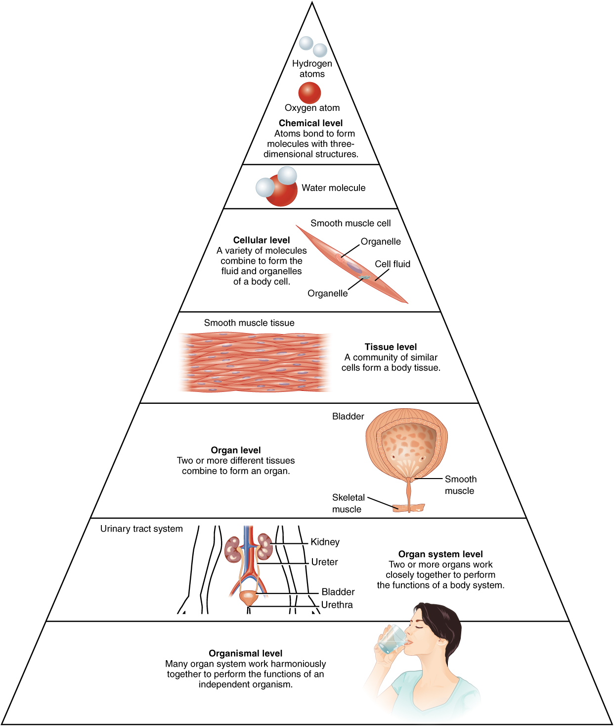
The organization of the human body can be seen as a hierarchy of increasing size and complexity, starting at the level of atoms and molecules, and ending at the level of the entire organism, which is an individual living thing. You can see the intervening levels of organization in Figure 7.2.2. Read about the levels in the sections that follow.
Figure 4.1 This diagram shows the levels of organization of the human body, from atoms to the whole organism.
CELLS
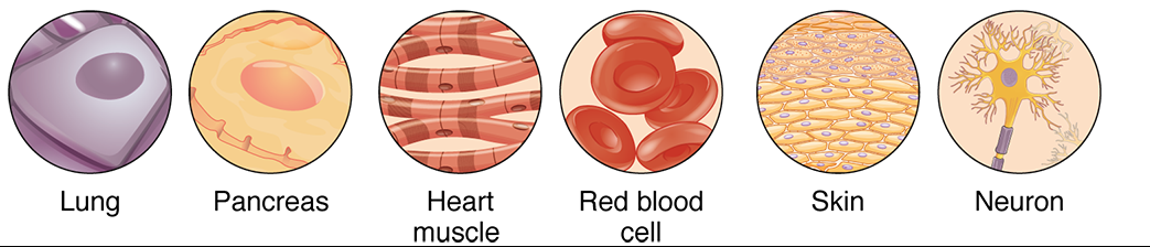
The basic units of structure and function of the human body — as in all living things — are cells. By the time the average person reaches adulthood, their body has an amazing 37 trillion of them! Each cell carries out basic life processes that allow the body to survive. In addition, most human cells are specialized in structure and function to carry out other specific roles. In fact, the human body may consist of as many as 200 different types of cells, each of which has a special job to do. Just a few of these different human cell types are pictured in Figure 4.2. These cells have obvious differences in structure that reflect their different functions. For example, nerve cells have long projections sticking out from the body of the cell. These projections help them carry electrical messages to other cells.
Figure 4.2 A few of the many different types of cells in the human body are illustrated here. Each type of cell is specialized for a particular role in the body.
TISSUES
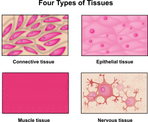
The next level of organization in the human body is tissues. A tissue is a group of connected cells that have a similar function. There are four basic types of human tissues: epithelial, muscle, nervous, and connective tissues. These four tissue types (shown in Figure 4.3) make up all the organs of the human body.
Figure 4.3 The human body contains these four types of tissues.
Organs and Organ Systems
Organs are the next level of organization of the human body. An organ is a structure that consists of two or more types of tissues that work together to do the same job. Examples of human organs include the heart, brain, lungs, skin, and kidneys. Human organs are organized into organ systems, which are shown in Figure 4.4. An organ system is a group of organs that work together to carry out a complex overall function. Each organ of the system does part of the larger job.
Figure 4.4 The Human Organ Systems. Some of the system names shown in this illustration differ from the terminology used in this book, but the systems are the same.
A Well-Oiled Machine
All of the organs and organ systems of the human body normally work together like a well-oiled machine, because they are closely regulated by the nervous and endocrine systems. The nervous system controls virtually all body activities, and the endocrine system secretes hormones that help to regulate these activities. Functioning together, the organ systems supply body cells with all the substances they need and eliminate their wastes. They also keep temperature, pH, and other conditions at just the right levels to support life.
Review
How is the human body like a complex machine?
Describe the difference between human anatomy and human physiology.
Relate cell structure to cell function, and give examples of specific cell types in the human body.
Define tissue, and identify the four types of tissues that make up the human body.
What is an organ? Give three examples of organs in the human body.
Define organ systems. Name five examples in the human body.
How is the human body regulated so all of its organs and organ systems work together?
4.3 TISSUES
Groups of connected cells form tissues. The cells in a tissue may all be the same type, or they may be of multiple types. In either case, the cells in the tissue work together to carry out a specific function. There are four main types of human tissues: connective, epithelial, muscle, and nervous tissues. We will be learning about each of the four tissue types in more detail in the next few sections, but here is a summary: The 200 types of cells that the body can produce based on our single set of DNA can create all the types of tissue in the body.
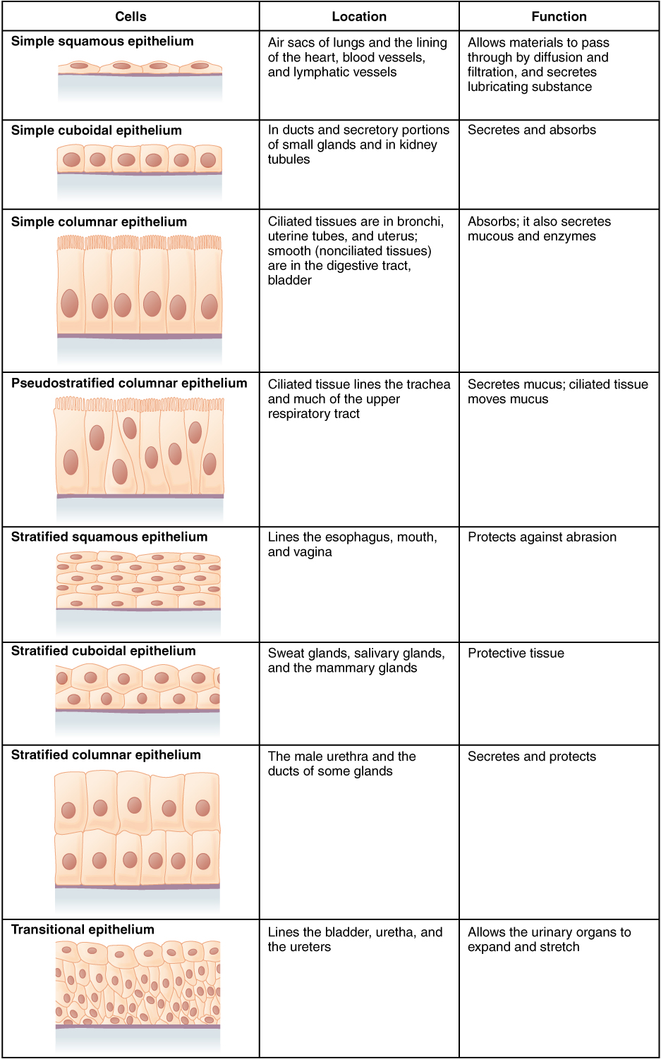
EPITHELIAL TISSUE
Epithelial tissue is made up of cells that line inner and outer body surfaces, such as the skin and the inner surface of the digestive tract. Epithelial tissue that lines inner body surfaces and body openings is called mucous membrane. This type of epithelial tissue produces mucus, a slimy substance that coats mucous membranes and traps pathogens, particles, and debris. Epithelial tissue protects the body and its internal organs, secretes substances (such as hormones) in addition to mucus, and absorbs substances (such as nutrients).
The key identifying feature of epithelial tissue is that it contains a free surface and a basement membrane. The free surface is not attached to any other cells and is either open to the outside of the body, or is open to the inside of a hollow organ or body tube. The basement membrane anchors the epithelial tissue to underlying cells.
Epithelial tissue is identified and named by shape and layering. Epithelial cells exist in three main shapes: squamous, cuboidal, and columnar. These specifically shaped cells can, depending on function, be layered in two ways: simple, stratified.
Epithelial tissue forms coverings and linings and is responsible for a range of functions including diffusion, absorption, secretion and protection. The shape of an epithelial cell can maximize its ability to perform a certain function. The thinner an epithelial cell is, the easier it is for substances to move through it to carry out diffusion and/or absorption. The larger an epithelial cell is, the more room it has in its cytoplasm to be able to make products for secretion, and the more protection it can provide for underlying tissues. Their are three main shapes of epithelial cells: squamous (which is shaped like a pancake- flat and oval), cuboidal (cube shaped), and columnar (tall and rectangular).
Epithelial tissue will also organize into different layering depending on their function. For example, multiple layers of cells provide excellent protection, but would no longer be efficient for diffusion, whereas a single layer would work very well for diffusion, but no longer be as protective.
Table 4.1 Summary of Epithelial Tissue Cells
Cell Junctions
Cells can communicate with each other via direct contact, referred to as intercellular junctions. Animal cell contacts include tight junctions, gap junctions, and desmosomes.
Tight Junctions
A tight junction is a watertight seal between two adjacent animal cells (Figure 3). The cells are held tightly against each other by proteins.
Figure 4.5. Tight junctions form watertight connections between adjacent animal cells. Proteins create tight junction adherence.
This tight adherence prevents materials from leaking between the cells; tight junctions are typically found in epithelial tissues that line internal organs and cavities and comprise most of the skin. For example, the tight junctions of the epithelial cells lining your urinary bladder prevent urine from leaking out into the extracellular space.
Desmosomes
Also found only in animal cells are desmosomes, which act like spot welds between adjacent epithelial cells (Figure 4.6). In the figure below, two adjacent cells are held together and the cells are maintained in a sheet-like formation in organs and tissues that stretch, like the skin, heart, and muscles.
Figure 4.6. A desmosome forms a very strong spot weld between cells. It is created by the linkage of cadherins and intermediate filaments. (credit: modification of work by Mariana Ruiz Villareal)
Gap Junctions
Gap junctions are channels between adjacent cells that allow for the transport of ions, nutrients, and other substances that enable cells to communicate.
Figure 4.7. A gap junction is a protein-lined pore that allows water and small molecules to pass between adjacent animal cells.
Gap junctions are particularly important in cardiac muscle: The electrical signal for the muscle to contract is passed efficiently through gap junctions, allowing the heart muscle cells to contract in tandem.
CONNECTIVE TISSUE
Bone and blood are examples of connective tissue. Connective tissue is very diverse. In general, it forms a framework and support structure for body tissues and organs. It’s made up of living cells separated by non-living material, called extracellular matrix which can be solid or liquid. The extracellular matrix of bone, for example, is a rigid mineral framework. The extracellular matrix of blood is liquid plasma.
The key identifying feature of connective tissue is that it is composed of a scattering of cells in a non-cellular matrix. There are two main categories of connective tissue: fibrous and special, based on the nature of the matrix.
FIBROUS CONNECTIVE TISSUE
Fibrous connective tissue contains cells called fibroblasts. These cells produce fibers of collagen, elastin, or reticular fiber which makes up the matrix of this type of connective tissue. Based on how tightly packed these fibers are and how they are oriented changes the properties, and therefore the function of the fibrous connective tissue.
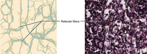
Reticular connective tissue is mostly composed of reticular protein fibers which make a skeleton, known as stroma, for the lymphatic and white blood cells. This type of tissue is found in the spleen and other lymphatic system structures.
Figure 4.8: Reticular Connective Tissue. This is a loose connective tissue made up of a network of reticular fibers that provides a supportive framework for soft organs
Elastic connective tissue: Made up of freely branching elastic fibers with fibroblasts in the spaces between the fibers, this tissue allows the kind of stretch that is found in the walls of arteries.
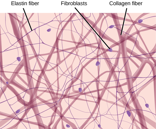
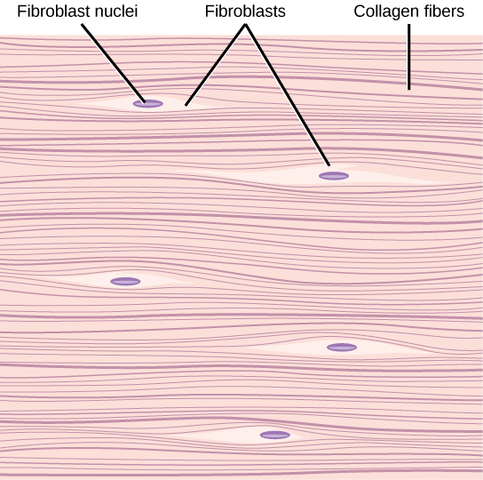
Loose fibrous connective tissue: composed of a loose and disorganized weave of collagen and elastin fibers, creating a tissue that is thin and flexible, yet still tough. This tissue, which is also sometimes referred to as “areolar tissue”, is found in membranes and surrounding blood vessels and most body organs. As you can see from the diagram in Figure 4.9, loose fibrous connective tissue fulfills the definition of connectives tissue since it is a scattering of cells (fibroblasts) in a non-cellular matrix (a mesh of collagen and elastin fibers). There are two types of specialized loose fibrous connective tissue: reticular and adipose. Adipose tissue stores fat and reticular tissue forms the spleen and lymph nodes.
Figure 4.9 Diagram of loose fibrous connective tissue consists of a scattering of fibroblasts in a non-cellular matrix of loosely woven collagen and elastin fibers.
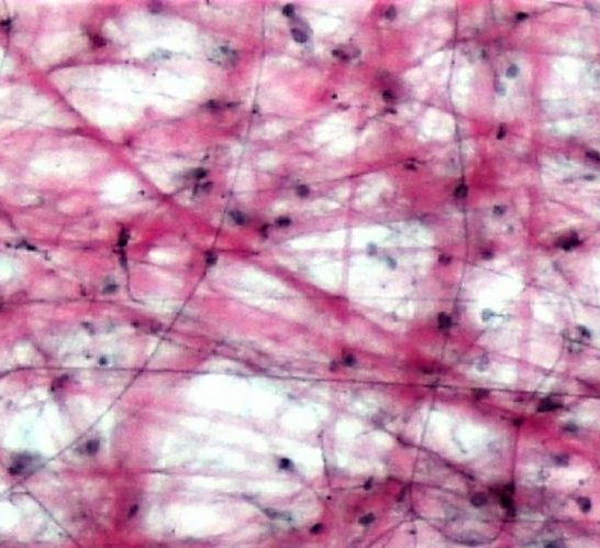
Figure 4.10 Microscopic view of loose fibrous connective tissue.
Dense Fibrous ConnectiveTissue: composed of a dense mat of parallel collagen fibers and a scattering of fibroblasts, creating a tissue that is very strong. Dense fibrous connective tissue forms tendons and ligaments, which connect bones to muscles and/or bones to neighboring bones.
Figure 4.11 Dense fibrous connective tissue is composed of fibroblasts and a dense parallel packing of collagen fibers.
Figure 4.12 Microscopic view of dense fibrous connective tissue.
SPECIAL CONNECTIVE TISSUE
Special connective tissue exhibits the defining feature of connective tissue in that it is a scattering of cells in a non-cellular matrix; what sets it apart from other connective tissues is its matrix. Some have solid matrices such as bone or cartilage. Blood is considered a connective tissue with a fluid matrix. While fibrous connective tissue contained cells called fibroblasts which produced fibers, solid connective tissue contains cells that either create bone (osteocytes) or cells that create cartilage (chondrocytes).
Cartilage
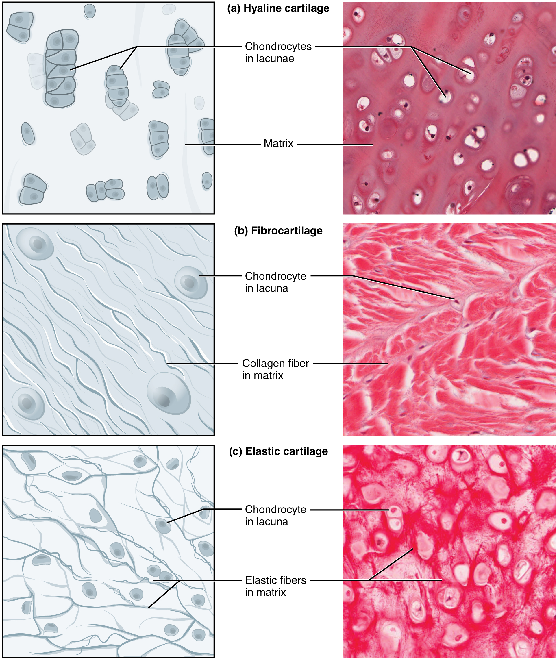
Chondrocytes produce the cartilage matrix in which they reside. Cartilage is made up of protein fibers and chondrocytes in lacunae. This is tissue is strong yet flexible and is used many places in the body for protection and support. Cartilage is one of the few tissues that is not vascular (doesn’t have a direct blood supply) meaning it relies on diffusion to obtain nutrients and gases; this is the cause of slow healing rates in injuries involving cartilage. There are three main types of cartilage:
Hyaline cartilage: a smooth, strong and flexible tissue. Found at the ends of ribs and long bones, in the nose, and comprising the entire fetal skeleton.
Fibrocartilage: a very strong tissue containing thick bundles of collagen. Found in joints that need cushioning from high impact (knees, jaw).
Elastic cartilage: contains elastic fibers in addition to collagen, giving support with the benefit of elasticity. Found in earlobes and the epiglottis. Figure 4.13: Three types of cartilage, each with distinct characteristics based on the nature of the matrix.
Bone
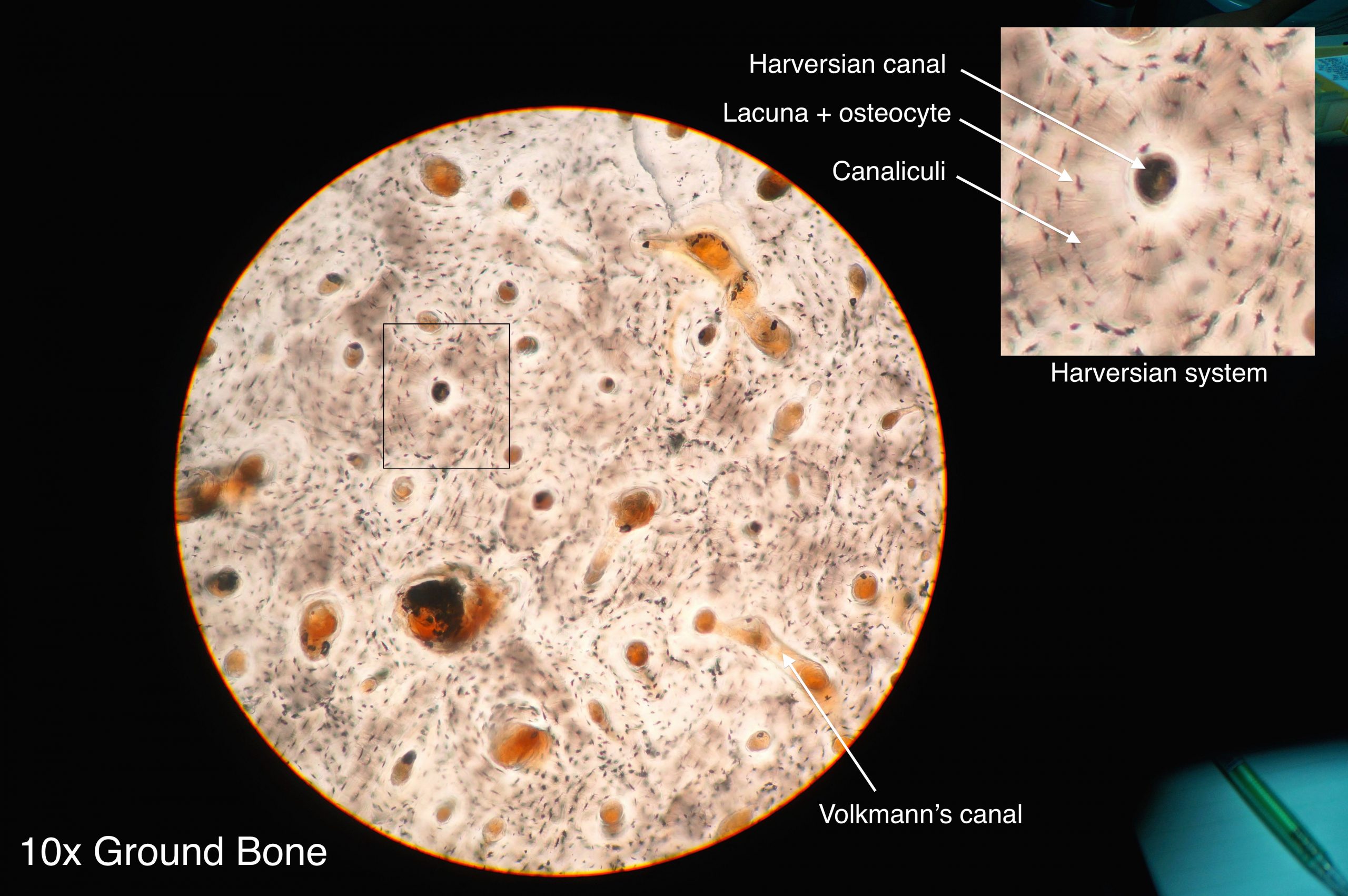
Osteocytes produce the bone matrix in which they reside. Since bone is very solid, these cells reside in small spaces called lacuna. This bone tissue is composed of collagen fibers embedded in calcium phosphate giving it strength without brittleness. There are two types of bone: compact and spongy.
Compact bone: has a dense matrix organized into cylindrical units called osteons. Each osteon contains a central canal (sometimes called a Harversian Canal) which allows for space for blood vessels and nerves, as well as concentric rings of bone matrix and osteocytes in lacunae, as per the diagram here. Compact bone is found in long bones and forms a shell around spongy bone.
Figure 4.14 Compact bone is composed of organized units called osteons.
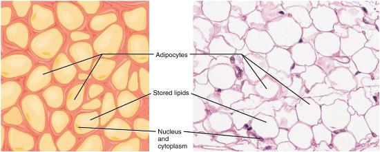
Adipose
Adipose connective tissue is commonly known as fat. This tissue contains fat cells that are specialized for lipid storage. In addition to storing energy, this tissue also cushions and protects the organs.
Figure 4.15: Adipose Connective Tissue consists of fat cells (adipocytes with a nucleus and stored lipids in their cytoplasm) with a little extracellular matrix. It stores fat for energy and provides insulation.
Blood
Blood has a matrix that is fluid; unlike the other two categories of connective tissue, the cells that reside in the matrix do not actually produce the matrix. Fibroblasts make the fibrous matrix, chondrocytes make the cartilaginous matrix, osteocytes make the bony matrix, yet blood cells do not make the fluid matrix of either lymph or plasma. This tissue still fits the definition of connective tissue in that it is still a scattering of cells in a non-cellular matrix.
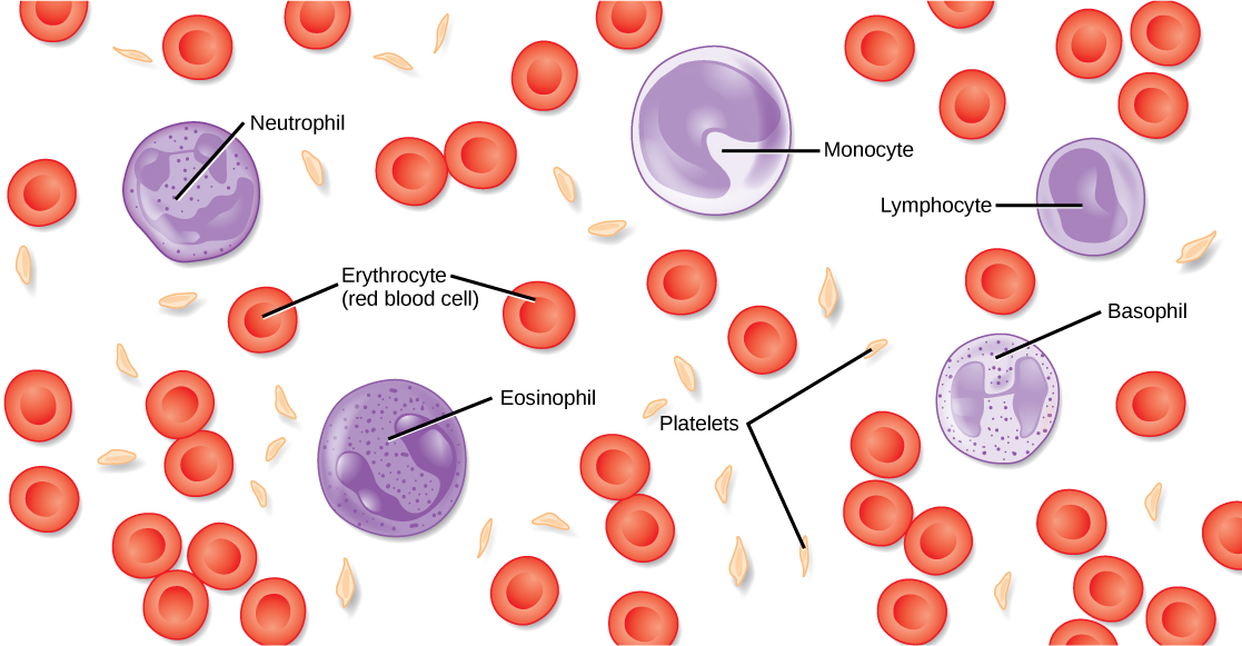
Blood: blood contains three types of cells suspended in plasma and is contained in the cardiovascular system.
Erythrocytes, more commonly called red blood cells, are present in high numbers (roughly 5 million cells per mL) and are responsible for delivering oxygen from to the lungs to all the other areas of the body. These cells are relatively small in size with a diameter of around 7 micrometers and live no longer than 120 days.
Leukocytes, often referred to as white blood cells, are present in lower numbers (approximately 5 thousand cells per mL) are responsible for various immune functions. They are typically larger than erythrocytes, but can live much longer, particularly white blood cells responsible for long term immunity. The number of leukocytes in your blood can go up or down based on whether or not you are fighting an infection.
Thrombocytes, also known as platelets, are very small cells responsible for blood clotting. Thrombocytes are not actually true cells; they are fragments of a much larger cell called a megakaryocyte.
Lymph: contains a liquid matrix and white blood cells and is contained in the lymphatic system, which ultimately drains into the cardiovascular system.
Figure 4.16: The cells and cellular components of human blood are shown. Red blood cells deliver oxygen to the cells and remove carbon dioxide. White blood cells (including neutrophils, monocytes, lymphocytes, eosinophils, and basophils) are involved in the immune response. Platelets form clots that prevent blood loss after injury.
MUSCULAR TISSUE
Muscular tissue is made up of cells that have the unique ability to contract- which is the defining feature of muscular tissue. There are three major types of muscle tissue.
SKELETAL MUSCLE
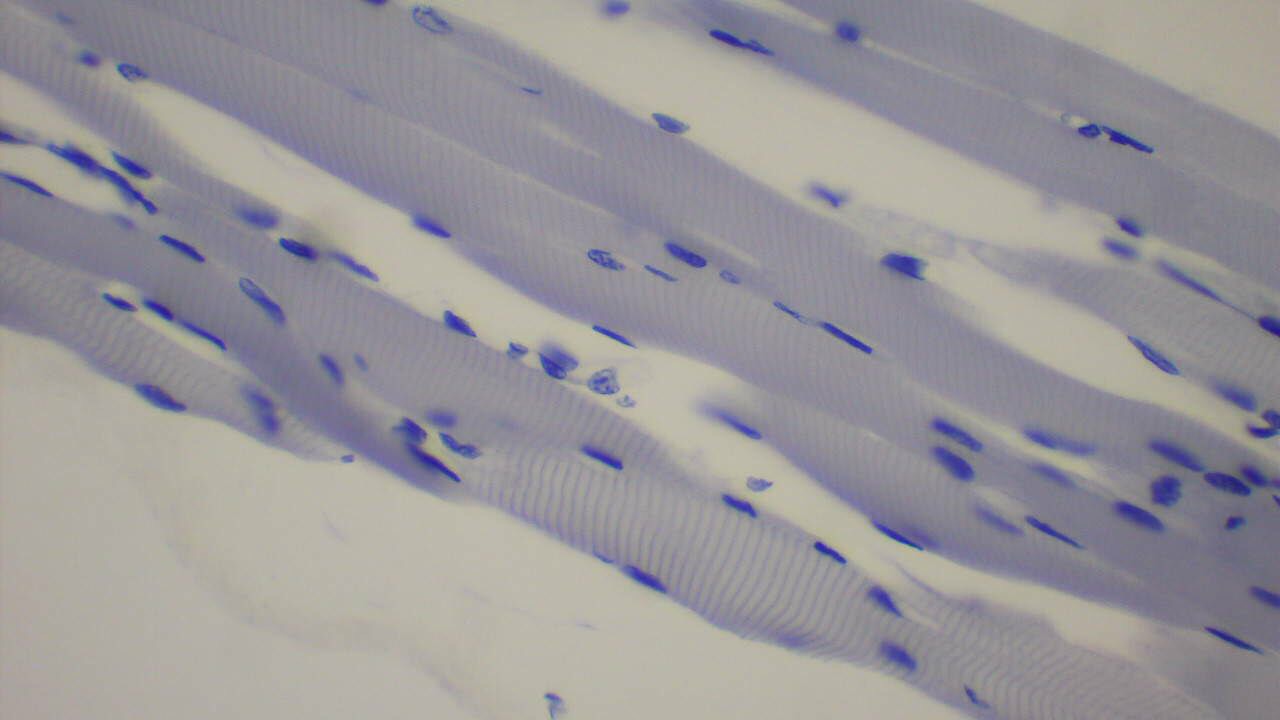
Skeletal muscles are voluntary muscles, meaning that you exercise conscious control over them. Skeletal muscles are attached to bones by tendons, a type of connective tissue. When these muscles shorten to pull on the bones to which they are attached, they enable the body to move. When you are exercising, reading a book, or making dinner, you are using skeletal muscles to move your body to carry out these tasks.
Under the microscope, skeletal muscles are striated (or striped) in appearance, because of their internal structure which contains alternating protein fibers of actin and myosin. Skeletal muscle is described as multinucleated, meaning one “cell” has many nuclei. This is because in utero, individual cells destined to become skeletal muscle fused, forming muscle fibers in a process known as myogenesis. You will learn more about skeletal muscle and how it contracts in the Muscular System.
Figure 4.17 Skeletal muscle is striated and multinucleated.
SMOOTH MUSCLE
Smooth muscles are nonstriated muscles- they still contain the muscle fibers actin and myosin, but not in the same alternating arrangement seen in skeletal muscle. Smooth muscle is found in the tubes of the body – in the walls of blood vessels and in the reproductive, gastrointestinal, and respiratory tracts. Smooth muscles are not under voluntary control meaning that they operate unconsciously, via the autonomic nervous system.
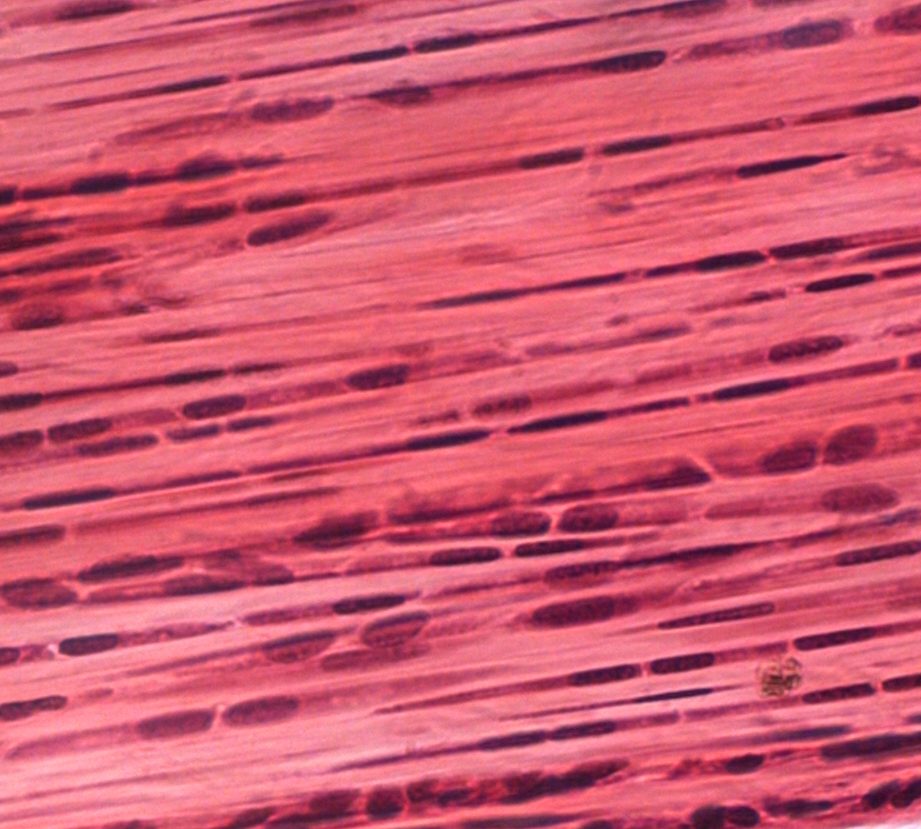
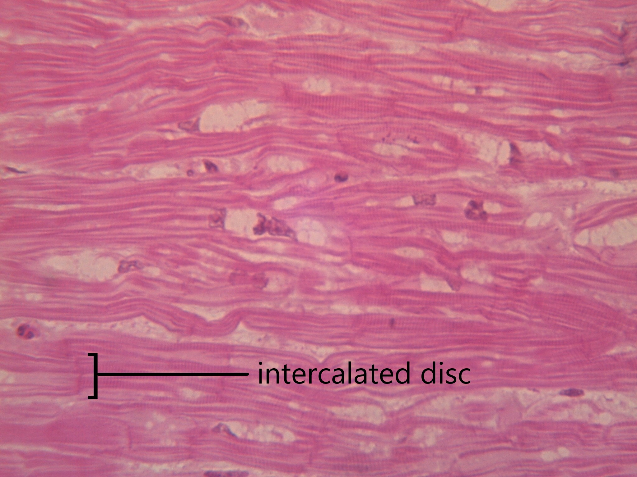
Cardiac muscles work involuntarily, meaning they are regulated by the autonomic nervous system. This is probably a good thing, since you wouldn’t want to have to consciously concentrate on keeping your heart beating all the time! Cardiac muscle, which is found only in the heart, is mononucleated and striated (due to alternating bands of myosin and actin). Their contractions cause the heart to pump blood. In order to make sure entire sections of the heart contract in unison, cardiac muscle tissue contains special cell junctions called intercalated discs, which conduct the electrical signals used to “tell” the chambers of the heart when to contract.
Figure 4.19 Cardiac muscle cells contain a single nucleus, have a striated appearance, and are joined by specialized junctions called intercalated discs.
NERVOUS TISSUE
Nervous tissue is made up of neurons and a group of cells called neuroglia (also known as glial cells). Nervous tissue makes up the central nervous system (mainly the brain and spinal cord) and peripheral nervous system (the network of nerves that runs throughout the rest of the body). The defining feature of nervous tissue is that it is specialized to be able to generate and conduct nerve impulses. This function is carried out by neurons, and the purpose of neuroglia is to support neurons.
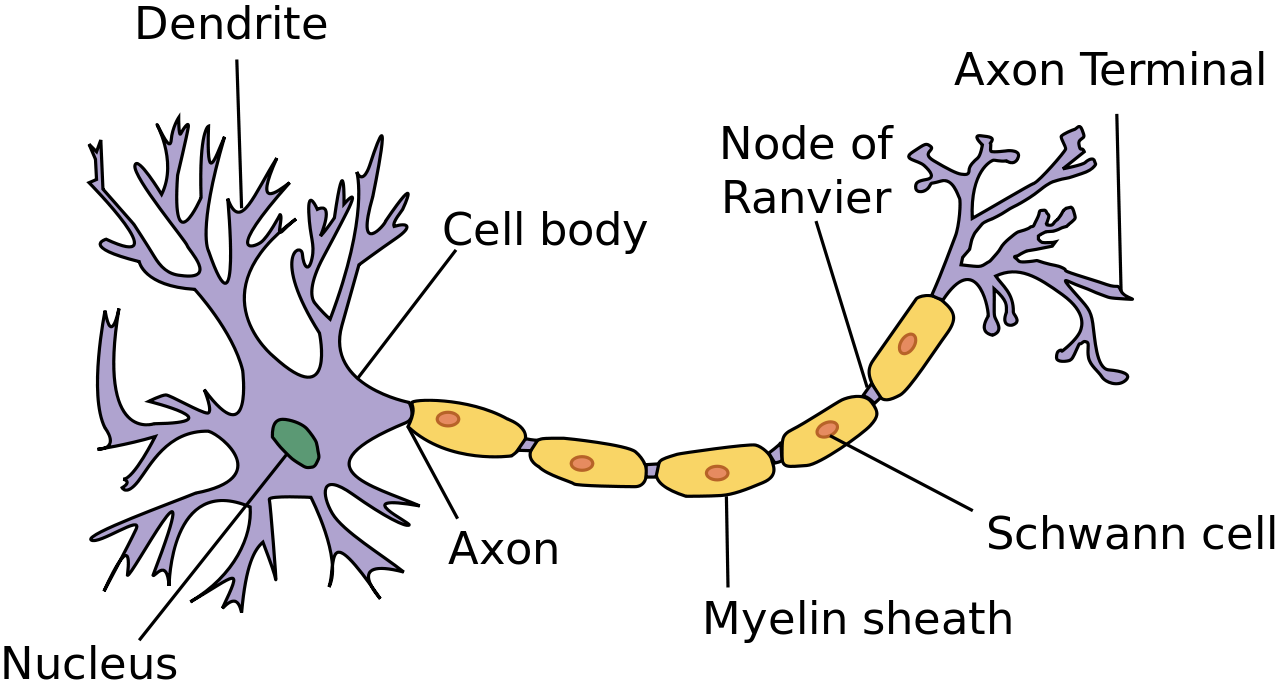
A neuron has several parts to its structure:
Dendrites which collect incoming nerve impulses
A cell body, or soma, which contains the majority of the neuron’s organelles, including the nucleus
An axon, which carries nerve impulses away from the soma to the next neuron in the chain
A myelin sheath, which encases the axon and increases that rate at which nerve impulses can be conducted
Axon terminals, which maintain physical contact with the dendrites of neighboring neurons
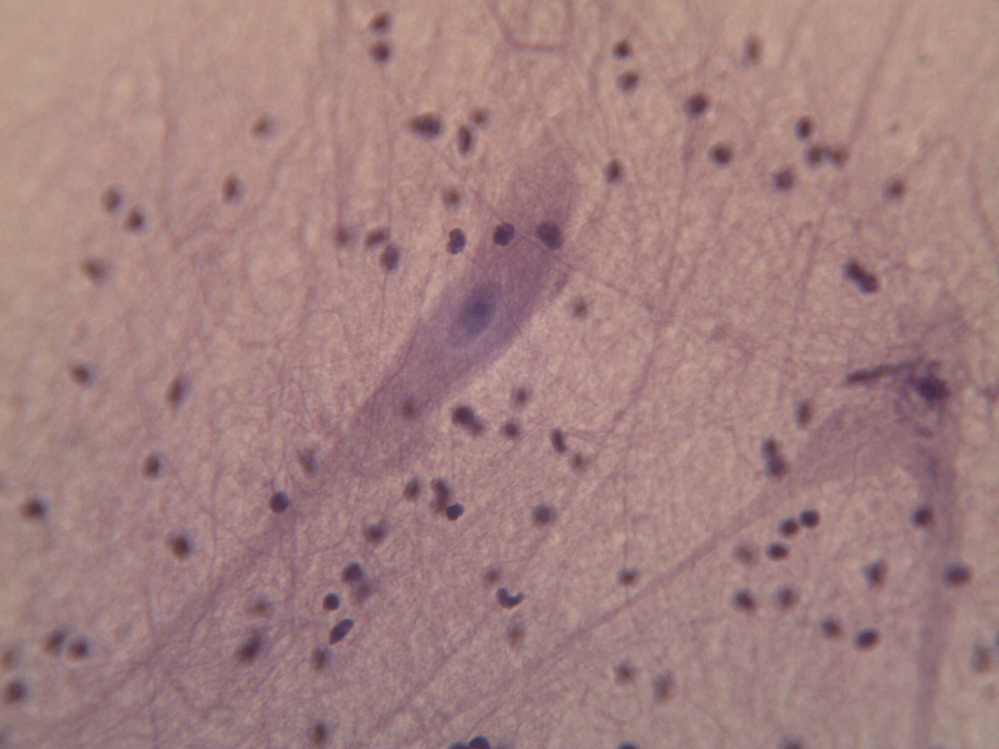
Neuroglia can be understood as support staff for the neuron. The neurons have such an important job, they need cells to bring them nutrients, take away cell waste, and build their myelin sheath. There are many types of neuroglia, which are categorized based on their function and/or their location in the nervous system. Neuroglia outnumber neurons by as much as 50 to 1 and are much smaller. See the diagram in 4.21 to compare the size and number of neurons and neuroglia.
Figure 4.20 Neurons a cell which specialize in conducting electrical impulses.
Figure 4.21 Neuroglia, the small cells seen here, outnumber neurons (the two larger cells) by as much as 50 to 1.
Review
Define the term tissue.
Fill in the missing words: Connective tissue is defined as containing a scattering of cells in a non-cellular _______________. The cells that reside in fibrous connective tissue are called ________________. The cells that lay down bone matrix are called ________. Blood has a _____________ matrix.
Where do you find skeletal muscle? Smooth muscle? Cardiac muscle?
What are the three types of cell junctions?
Fill in the blanks for the following:
Epithelial tissue makes up the linings and ________________ of the body.
Epithelial tissue cells can come in three shapes: squamous, cuboidal, and ________________. These cells also vary in how they are layered: a single layer of cells is termed _______________ and many layers are termed ________________.
Your skin is an epithelial tissue designed for _________________ due to its many layers. The simple squamous epithelial tissue in your lungs is designed for __________________..Word Bank:
stratified
diffusion
protection
simple
coverings
columnar
What type of tissue is pictured below?
4.4 HUMAN ORGAN SYSTEMS
Functionally related organs often cooperate to form whole organ systems. The 12 diagrams in Figure 4.4 above show 11 human organ systems, including separate diagrams for the male and female reproductive systems. Some of the organs and functions of the organ systems are identified. Each system is also described in more detail in the text that follows. Most of these human organ systems are also the subject of separate chapters in this book.
INTEGUMENTARY SYSTEM
Organs of the integumentary system include the skin, hair, and nails. The skin is the largest organ in the body. It encloses and protects the body and is the site of many sensory receptors. The skin is the body’s first defense against pathogens, and it also helps regulate body temperature and eliminate wastes in sweat.
SKELETAL SYSTEM
The skeletal system consists of bones, joints, teeth. The bones of the skeletal system are connected by tendons, ligaments, and cartilage. Functions of the skeletal system include supporting the body and giving it shape. Along with the muscular system, the skeletal system enables the body to move. The bones of the skeletal system also protect internal organs, store calcium, and produce red and white blood cells.
MUSCULAR SYSTEM
The muscular system consists of three different types of muscles, including skeletal muscles, which are attached to bones by tendons and allow for voluntary movements of the body. Smooth muscle tissues control the involuntary movements of internal organs, such as the organs of the digestive system, allowing food to move through the system. Smooth muscles in blood vessels allow vasoconstriction and vasodilation, thereby helping to regulate body temperature. Cardiac muscle tissues control the involuntary beating of the heart, allowing it to pump blood through the blood vessels of the cardiovascular system.
NERVOUS SYSTEM
The nervous system includes the brain and spinal cord — which make up the central nervous system — and nerves that run throughout the rest of the body, making up the peripheral nervous system. The nervous system controls both voluntary and involuntary responses of the human organism, and also detects and processes sensory information.
ENDOCRINE SYSTEM
The endocrine system is made up of glands that secrete hormones into the blood, which then carries hormones throughout the body. Endocrine hormones are chemical messengers that control many body functions, including metabolism, growth, and sexual development. The master gland of the endocrine system is the pituitary gland, which produces hormones that control other endocrine glands. Some of the other endocrine glands include the pancreas, thyroid gland, and adrenal glands.
CARDIOVASCULAR SYSTEM
The cardiovascular system (also called circulatory system) includes the heart, blood, and three types of blood vessels: arteries, veins, and capillaries. The heart pumps blood, which travels through the blood vessels. The main function of the cardiovascular system is transport. Oxygen from the lungs and nutrients from the digestive system are transported to cells throughout the body. Carbon dioxide and other waste materials are picked up from the cells and transported to organs (such as the lungs and kidneys) for elimination from the body. The cardiovascular system also equalizes body temperature and transports endocrine hormones to cells in the body where they are needed.
LYMPHATIC SYSTEM
The lymphatic system is sometimes considered part of the immune system. It consists of a network of lymph vessels and ducts that collect excess fluid (called lymph) from extracellular spaces in tissues and transport the fluid to the bloodstream. The lymphatic system also includes many small collections of tissue, (called lymph nodes) and an organ called the spleen, both of which remove pathogens and cellular debris from the lymph or blood. In addition, the thymus gland in the lymphatic system produces some types of white blood cells (lymphocytes) that fight infections.
RESPIRATORY SYSTEM
Organs and other structures of the respiratory system include the nasal passages, lungs, and a long tube called the trachea, which carries air between the nasal passages and lungs. The main function of the respiratory system is to deliver oxygen to the blood and remove carbon dioxide from the body. Gases are exchanged between the lungs and blood across the walls of capillaries lining tiny air sacs (alveoli) in the lungs.
DIGESTIVE SYSTEM
The digestive system consists of several main organs — including the mouth, esophagus, stomach, and small and large intestines — that form a long tube called the gastrointestinal (GI) tract. Food moves through this tract, where it is digested. Its nutrients are then absorbed, and its waste products are excreted. The digestive system also includes accessory organs (such as the pancreas and liver) that produce enzymes and other substances needed for digestion, but through which food does not actually pass.
URINARY SYSTEM
The urinary system is part of the excretory system, which removes wastes from the body. The urinary system includes the pair of kidneys, which filter excess water and a waste product (called urea) from the blood and form urine. Two tubes called ureters carry the urine from the kidneys to the urinary bladder, which stores the urine until it is excreted from the body through another tube called the urethra. The kidneys also produce an enzyme called renin and a variety of hormones. These substances help regulate blood pressure, the production of red blood cells, and the balance of calcium and phosphorus in the body.
MALE AND FEMALE REPRODUCTIVE SYSTEMS
The reproductive system is the only body system that differs substantially between males and females. Both male and female reproductive systems produce sex-specific sex hormones (testosterone in males, estrogen in females) and gametes (sperm in males, eggs in females). However, the organs involved in these processes are different. The male reproductive system includes the epididymis, testes, and penis. The female reproductive system includes the uterus, ovaries, and mammary glands. The male and female systems also have different additional roles. For example, the male system has the role of delivering gametes to the female reproductive tract, whereas the female system has the roles of supporting an embryo and fetus until birth and also producing milk for the infant after birth.
Human Body 101 | National Geographic, 2017.
4.5 BODY CAVITIES
What Are Body Cavities?
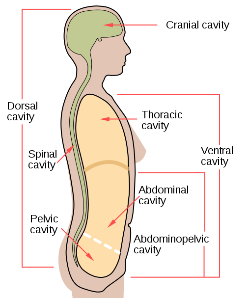
The human body, like that of many other multicellular organisms, is divided into a number of body cavities. A body cavity is a fluid-filled space inside the body that holds and protects internal organs. Human body cavities are separated by membranes and other structures. The two largest human body cavities are the ventral cavity and dorsal cavity. These two body cavities are subdivided into smaller body cavities. Both the dorsal and ventral cavities and their subdivisions are shown in the Figure 4.22 diagram.
Figure 4.22 Human body cavities.
Ventral Cavity
The ventral cavity is at the anterior (or front) of the trunk. Organs contained within this body cavity include the lungs, heart, stomach, intestines, and reproductive organs. The ventral cavity allows for considerable changes in the size and shape of the organs inside as they perform their functions. Organs such as the lungs, stomach, or uterus, for example, can expand or contract without distorting other tissues or disrupting the activities of nearby organs.
The thoracic cavity fills the chest and is subdivided into two pleural cavities and the pericardial cavity. The pleural cavities hold the lungs, and the pericardial cavity holds the heart.
The abdominopelvic cavity fills the lower half of the trunk and is subdivided into the abdominal cavity and the pelvic cavity. The abdominal cavity holds digestive organs and the kidneys, and the pelvic cavity holds reproductive organs and organs of excretion.
Dorsal Cavity
The dorsal cavity is at the posterior (or back) of the body, including both the head and the back of the trunk. The dorsal cavity is subdivided into the cranial and spinal cavities.
The cranial cavity fills most of the upper part of the skull and contains the brain.
The spinal cavity is a very long, narrow cavity inside the vertebral column. It runs the length of the trunk and contains the spinal cord.
The brain and spinal cord are protected by the bones of the skull and the vertebrae of the spine. They are further protected by the meninges, a three-layer membrane that encloses the brain and spinal cord. A thin layer of cerebrospinal fluid is maintained between two of the meningeal layers. This clear fluid is produced by the brain, and it provides extra protection and cushioning for the brain and spinal cord.
Feature: My Human Body
The meninges membranes that protect the brain and spinal cord inside their cavities may become inflamed, generally due to a bacterial or viral infection. This condition is called meningitis, and it can lead to serious long-term consequences such as deafness, epilepsy, or cognitive deficits, especially if not treated quickly. Meningitis can also rapidly become life-threatening, so it is classified as a medical emergency.
Learning the symptoms of meningitis may help you or a loved one get prompt medical attention if you ever develop the disease. Common symptoms include fever, headache, and neck stiffness. Other symptoms include confusion or altered consciousness, vomiting, and an inability to tolerate light or loud noises. Young children often exhibit less specific symptoms, such as irritability, drowsiness, or poor feeding.
Meningitis is diagnosed with a lumbar puncture (commonly known as a “spinal tap”), in which a needle is inserted into the spinal canal to collect a sample of cerebrospinal fluid. The fluid is analyzed in a medical lab for the presence of pathogens. If meningitis is diagnosed, treatment consists of antibiotics and sometimes antiviral drugs. Corticosteroids may also be administered to reduce inflammation and the risk of complications (such as brain damage). Supportive measures such as IV fluids may also be provided.
Some types of meningitis can be prevented with a vaccine. Ask your health care professional whether you have had the vaccine or should get it. Giving antibiotics to people who have had significant exposure to certain types of meningitis may reduce their risk of developing the disease. If someone you know is diagnosed with meningitis and you are concerned about contracting the disease yourself, see your doctor for advice.
Review
What is a body cavity?
Compare and contrast the ventral and dorsal body cavities.
Identify the subdivisions of the ventral cavity, and the organs each contains.
Describe the subdivisions of the dorsal cavity and their contents.
4.6 HOMEOSTASIS AND FEEDBACK
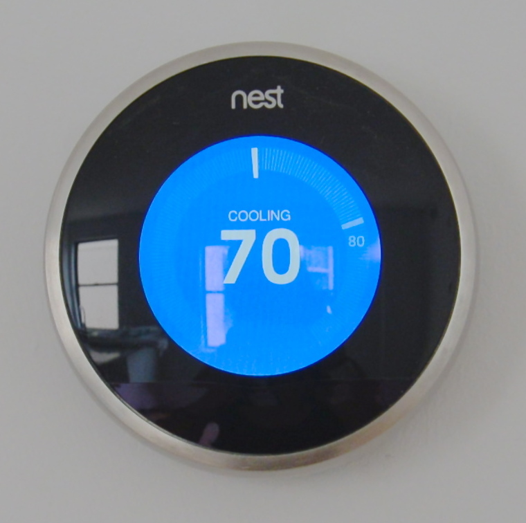
Figure 4.23 A thermostat controls a complex system to maintain a steady temperature in our homes.
STEADY AS SHE GOES
This device (Figure 4.23) looks simple, but it controls a complex system that keeps a home at a steady temperature — it’s a thermostat. The device shows the current temperature in the room, and also allows the occupant to set the thermostat to the desired temperature. A thermostat is a commonly cited model of how living systems — including the human body— maintain a steady state called homeostasis.
WHAT IS HOMEOSTASIS?
Homeostasis is the condition in which a system (such as the human body) is maintained in a more or less steady state. It is the job of cells, tissues, organs, and organ systems throughout the body to maintain many different variables within narrow ranges compatible with life. Keeping a stable internal environment requires continually monitoring the internal environment and constantly making adjustments to keep things in balance.
SET POINT AND NORMAL RANGE
For any given variable, such as body temperature or blood glucose level, there is a particular set point that is the physiological optimum value. The set point for human body temperature, for example, is about 37 degrees C (98.6 degrees F). As the body works to maintain homeostasis for temperature or any other internal variable, the value typically fluctuates around the set point. Such fluctuations are normal, as long as they do not become too extreme. The spread of values within which such fluctuations are considered insignificant is called the normal range. In the case of body temperature, for example, the normal range for an adult is about 36.5 to 37.5 degrees C (97.7 to 99.5 degrees F).
A good analogy for set point, normal range, and maintenance of homeostasis is driving. When you are driving a vehicle on the road, you are supposed to drive in the center of your lane — this is analogous to the set point. Sometimes, you are not driving in the exact center of the lane, but you are still within your lines, so you are in the equivalent of the normal range. However, if you were to get too close to the center line or the shoulder of the road, you would take action to correct your position. You’d move left if you were too close to the shoulder, or right if too close to the center line — which is analogous to our next concept, negative feedback to maintain homeostasis.
MAINTAINING HOMEOSTASIS
Homeostasis is normally maintained in the human body by an extremely complex balancing act. Regardless of the variable being kept within its normal range, maintaining homeostasis requires at least four interacting components: stimulus, sensor, control center, and effector.
The stimulus is provided by the variable being regulated. Generally, the stimulus indicates that the value of the variable has moved away from the set point or has left the normal range.
The sensor monitors the values of the variable and sends data on it to the control center.
The control centre matches the data with normal values. If the value is not at the set point or is outside the normal range, the control center sends a signal to the effector.
The effector is an organ, gland, muscle, or other structure that acts on the signal from the control center to move the variable back toward the set point.
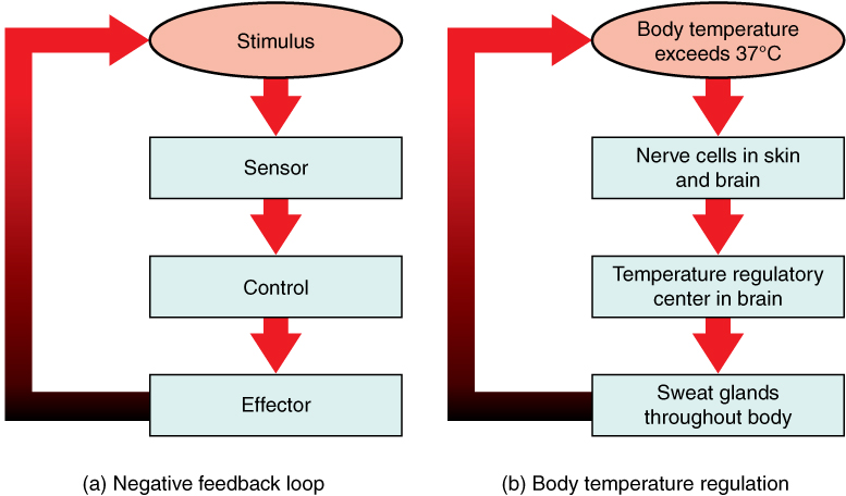
Each of these components is illustrated in Figure 4.24. The diagram on the left is a general model showing how the components interact to maintain homeostasis. The diagram on the right shows the example of body temperature. From the diagrams, you can see that maintaining homeostasis involves feedback, which is data that feeds back to control a response. Feedback may be negative (as in the example below) or positive. All the feedback mechanisms that maintain homeostasis use negative feedback. Biological examples of positive feedback are much less common.
Figure 4.24 Maintaining homeostasis through feedback requires a stimulus, sensor, control centre, and effector.
Negative Feedback
In a negative feedback loop, feedback serves to reduce an excessive response and keep a variable within the normal range. Two processes controlled by negative feedback are body temperature regulation and control of blood glucose.
BODY TEMPERATURE
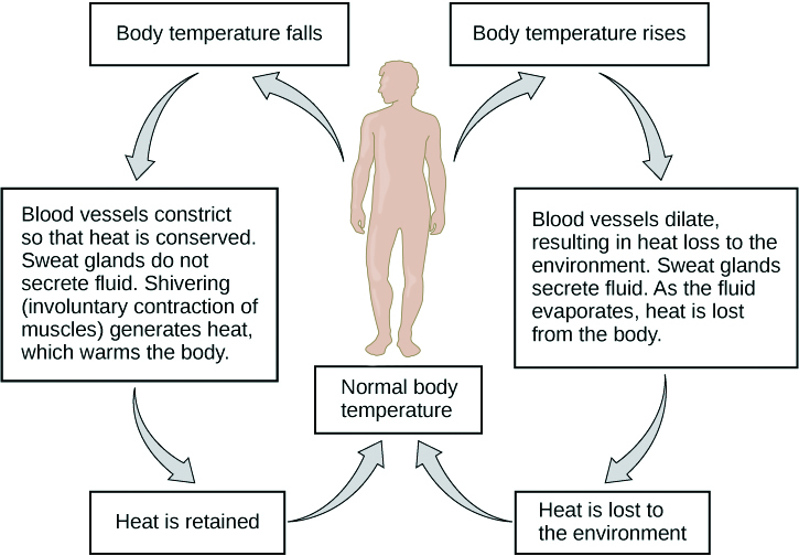
Body temperature regulation involves negative feedback, whether it lowers the temperature or raises it, as shown in Figure 4.25 and explained in the text that follows.
Figure 4.25 Homeostasis of body temperature is maintained by negative feedback loops.
Cooling Down
The human body’s temperature regulatory center is the hypothalamus in the brain. When the hypothalamus receives data from sensors in the skin and brain that body temperature is higher than the set point, it sets into motion the following responses:
Blood vessels in the skin dilate (vasodilation) to allow more blood from the warm body core to flow close to the surface of the body, so heat can be radiated into the environment.
As blood flow to the skin increases, sweat glands in the skin are activated to increase their output of sweat (diaphoresis). When the sweat evaporates from the skin surface into the surrounding air, it takes heat with it.
Breathing becomes deeper, and the person may breathe through the mouth instead of the nasal passages. This increases heat loss from the lungs.
Heating Up
When the brain’s temperature regulatory center receives data that body temperature is lower than the set point, it sets into motion the following responses:
Blood vessels in the skin contract (vasoconstriction) to prevent blood from flowing close to the surface of the body, which reduces heat loss from the surface.
As temperature falls lower, random signals to skeletal muscles are triggered, causing them to contract. This causes shivering, which generates a small amount of heat.
The thyroid gland may be stimulated by the brain (via the pituitary gland) to secrete more thyroid hormone. This hormone increases metabolic activity and heat production in cells throughout the body.
The adrenal glands may also be stimulated to secrete the hormone adrenaline. This hormone causes the breakdown of glycogen (the carbohydrate used for energy storage in animals) to glucose, which can be used as an energy source. This catabolic chemical process is exothermic, or heat producing.
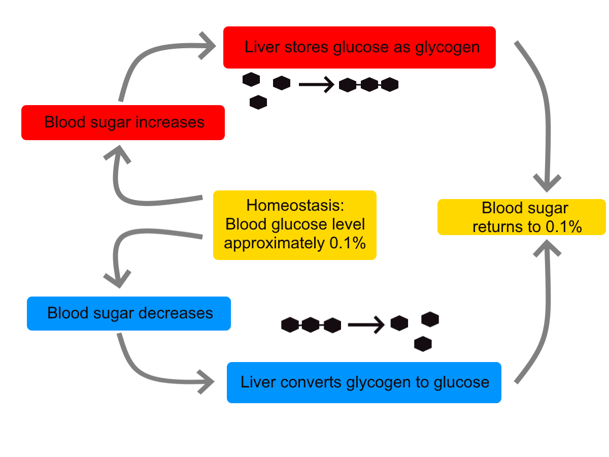
BLOOD GLUCOSE
In controlling the blood glucose level, certain endocrine cells in the pancreas (called alpha and beta cells) detect the level of glucose in the blood. They then respond appropriately to keep the level of blood glucose within the normal range.
If the blood glucose level rises above the normal range, pancreatic beta cells release the hormone insulin into the bloodstream. Insulin signals cells to take up the excess glucose from the blood until the level of blood glucose decreases to the normal range.
If the blood glucose level falls below the normal range, pancreatic alpha cells release the hormone glucagon into the bloodstream. Glucagon signals cells to break down stored glycogen to glucose and release the glucose into the blood until the level of blood glucose increases to the normal range.
Figure 4.26 Your liver plays an important role in balancing blood sugar levels. Glycogen in your liver can either collect glucose out of your blood stream to lower blood sugar, or release glucose into the bloodstream to increase blood sugar. This happens through a negative feedback loop.
POSITIVE FEEDBACK
In a positive feedback loop, feedback serves to intensify a response until an end point is reached. Examples of processes controlled by positive feedback in the human body include blood clotting and childbirth.
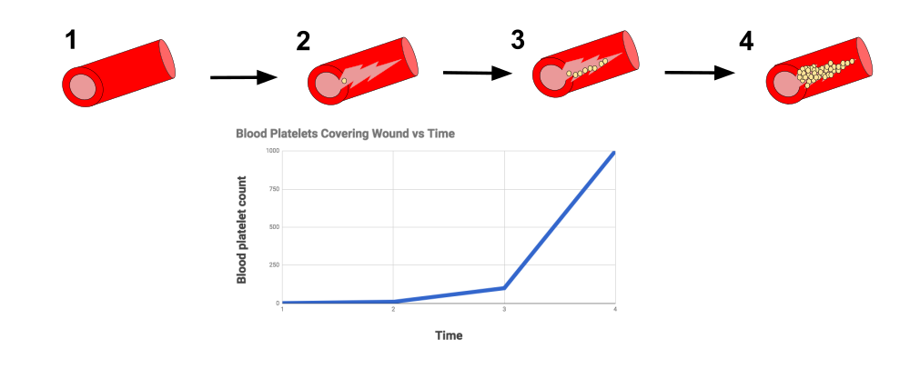
BLOOD CLOTTING
Figure 4.27 The diagram demonstrates positive feedback, using the example of blood clotting in the body. The damaged blood vessel wall releases chemicals that initiate the formation of a blood clot. Every time the blood clot builds up more, more chemicals are released that speed up the process. The process gets faster and faster until the blood vessel wall is completely healed and the positive feedback loop has ended. The graph represents the number of platelets aiding in the formation of the blood clot. The exponential form of the graph represents the positive feedback mechanism.
When a wound causes bleeding, the body responds with a positive feedback loop to clot the blood and stop blood loss. Substances released by the injured blood vessel wall begin the process of blood clotting. Platelets in the blood start to cling to the injured site and release chemicals that attract additional platelets. As the platelets continue to amass, more of the chemicals are released and more platelets are attracted to the site of the clot. The positive feedback accelerates the process of clotting until the clot is large enough to stop the bleeding.
CHILDBIRTH
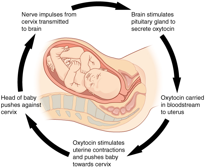
Figure 4.28 shows the positive feedback loop that controls childbirth. The process normally begins when the head of the infant pushes against the cervix. This stimulates nerve impulses, which travel from the cervix to the hypothalamus in the brain. In response, the hypothalamus sends the hormone oxytocin to the pituitary gland, which secretes it into the bloodstream so it can be carried to the uterus. Oxytocin stimulates uterine contractions, which push the baby harder against the cervix. In response, the cervix starts to dilate in preparation for the passage of the baby. This cycle of positive feedback continues, with increasing levels of oxytocin, stronger uterine contractions, and wider dilation of the cervix until the baby is pushed through the birth canal and out of the body. At that point, the cervix is no longer stimulated to send nerve impulses to the brain, and the entire process stops.
Figure 4.28 Normal childbirth is driven by a positive feedback loop.
Normal childbirth is driven by a positive feedback loop. Positive feedback causes an increasing deviation from the normal state to a fixed end point, rather than a return to a normal set point as in homeostasis.
WHEN HOMEOSTASIS FAILS
Homeostatic mechanisms work continuously to maintain stable conditions in the human body. Sometimes, however, the mechanisms fail. When they do, homeostatic imbalance may result, in which cells may not get everything they need or toxic wastes may accumulate in the body. If homeostasis is not restored, the imbalance may lead to disease — or even death. Diabetes is an example of a disease caused by homeostatic imbalance. In the case of diabetes, blood glucose levels are no longer regulated and may be dangerously high. Medical intervention can help restore homeostasis and possibly prevent permanent damage to the organism.
Normal aging may bring about a reduction in the efficiency of the body’s control systems, which makes the body more susceptible to disease. Older people, for example, may have a harder time regulating their body temperature. This is one reason they are more likely than younger people to develop serious heat-induced illnesses, such as heat stroke.
GCSE Biology – Homeostasis #38, Cognito, 2018.
Review
Compare and contrast negative and positive feedback loops.
Explain how negative feedback controls body temperature.
Give two examples of physiological processes controlled by positive feedback loops.
During breastfeeding, the stimulus of the baby sucking on the nipple increases the amount of milk produced by the mother. The more sucking, the more milk is usually produced. Is this an example of negative or positive feedback? Explain your answer. What do you think might be the evolutionary benefit of the milk production regulation mechanism you described?
Explain why homeostasis is regulated by negative feedback loops, rather than positive feedback loops.
4.7 CASE STUDY: UNDER PRESSURE
Figure 4.29 Football often involves forceful impact to the head which makes wearing a helmet critical.
Looking at this photo of a football game (Figure 4.29), you can see why it is so important that the players wear helmets. As players tackle each other, football often involves forceful impact to the head. This can cause damage to the brain — temporarily (as in the case of a concussion) or long-term and more severe. Helmets are critical in reducing the incidence of traumatic brain injuries (TBIs), but they do not fully prevent them.
As a former professional football player who also played in college and high school, 43-year-old Jayson sustained many high-impact head injuries over the course of his football playing years. A few years ago, Jayson began experiencing a variety of troubling symptoms, including the loss of bladder control (the involuntary leakage of urine), memory loss, and difficulty walking. Symptoms like these are often signs of damage to the nervous system, which includes the brain, spinal cord, and nerves, but they can result from many different types of injuries or diseases that affect the nervous system. In order to treat him properly, Jayson’s doctors needed to do several tests to determine the exact cause of his symptoms. The doctors ordered a spinal tap to see if he had an infection, and an MRI (magnetic resonance imaging) to see if there were any observable problems in and around his brain.
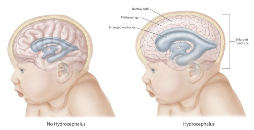
The MRI revealed the cause of Jayson’s symptoms. There are fluid-filled spaces within the brain called ventricles, and compared to normal ventricles, Jayson’s ventricles were enlarged. Based on this observation, along with the results of other tests, Jayson’s doctor diagnosed him with hydrocephalus, a term that literally means “water head.” Hydrocephalus occurs when the fluid that fills the ventricles — called cerebrospinal fluid — builds up excessively, causing the ventricles to become enlarged. This puts pressure on the brain, which can cause a variety of neurological symptoms, including the ones Jayson was experiencing. In Figure 4.30, you can see the difference between normal ventricles and ventricles that are enlarged due to hydrocephalus. Notice in the image on the right how the brain becomes “squeezed” due to hydrocephalus.
Figure 4.30Comparison of an infant with and without hydrocephalus. The ventricles (shown in blue gray) are located inside the brain (shown in pink).
Hydrocephalus often occurs at birth, as a result of genetic factors or events that occurred during fetal development. Because babies are born with skull bones that are not fully fused, the skull of a baby born with hydrocephalus can expand and relieve some of the pressure on the brain, as reflected in the enlarged head size shown in Figure 7.1.2. Adults have fully fused, inflexible skulls, so when hydrocephalus occurs in an adult, the brain experiences all of the increased pressure.
Why did Jayson develop hydrocephalus? There are many possible causes of hydrocephalus in adults, including tumors, infections, hemorrhages, and TBIs. Given his repeated and long history of football-related TBIs and the absence of any evidence of infection, tumor, or other cause, Jayson’s doctor thinks his head injuries were most likely responsible for his hydrocephalus.
Although hydrocephalus is serious, there are treatments. Read the rest of this chapter to learn about the cells, tissues, organs, cavities, and systems of the body, how they are interconnected, and the importance of keeping the body in a state of homeostasis (or balance). The amount of cerebrospinal fluid in the ventricles is normally kept at a relatively steady level, and the potentially devastating symptoms of hydrocephalus are an example of what can happen when a system in the body becomes unbalanced. At the end of the chapter, you will learn about Jayson’s treatment and prognosis.
As you learned in this chapter, the human body consists of many complex systems that normally work together efficiently — like a well-oiled machine — to carry out life’s functions. The brain and spinal cord are protected by layers of membrane called meninges and fluid that flows between the meninges and in spaces called ventricles inside the brain. This fluid is called cerebrospinal fluid, and as you have learned, one of its important functions is to cushion and protect the brain and spinal cord, which make up most of the central nervous system (CNS). Additionally, cerebrospinal fluid circulates nutrients and removes waste products from the CNS. Cerebrospinal fluid is produced continually in the ventricles, circulates throughout the CNS, and is then reabsorbed by the bloodstream. If too much cerebrospinal fluid is produced, its flow is blocked, or not enough is reabsorbed, the system becomes out of balance and it can build up in the ventricles. This causes an enlargement of the ventricles called hydrocephalus that can put pressure on the brain, resulting in the types of neurological problems that former professional football player Jayson, described in the beginning of this chapter, is suffering from.
Jayson’s symptoms included loss of bladder control, memory loss, and difficulty walking. The cause of his symptoms was not immediately clear, although his doctors suspected that it related to the nervous system, since the nervous system acts as the control center of the body, controlling and regulating many other organ systems. Jayson’s memory loss directly implicated the brain’s involvement, since that is the site of thoughts and memory. The urinary system is also controlled in part by the nervous system, so the inability to hold urine appropriately can also be a sign of a neurological issue. Jayson’s trouble walking involved the muscular system, which works alongside the skeletal system to enable movement of the limbs. In turn, the contraction of muscles is regulated by the nervous system. You can see why a problem in the nervous system can cause a variety of different symptoms by affecting multiple organ systems in the human body.
To try to find the exact cause of Jayson’s symptoms, his doctors performed a lumbar puncture (or spinal tap), which is the removal of some cerebrospinal fluid through a needle inserted into the lower part of the spinal canal. They then analyzed Jayson’s cerebrospinal fluid for the presence of pathogens (such as bacteria) to determine whether an infection was the cause of his neurological symptoms. When no evidence of infection was found, they used an MRI to observe the structures of his brain. This is when they discovered his enlarged ventricles, which are a hallmark of hydrocephalus.
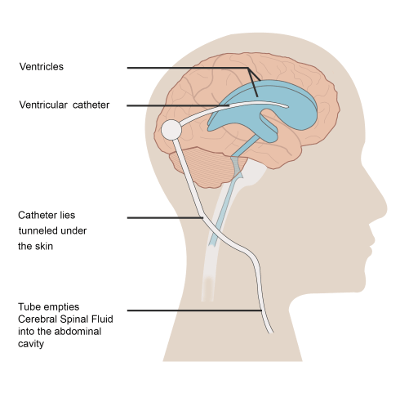
To treat Jayson’s hydrocephalus, a surgeon implanted a device called a shunt in his brain to remove the excess fluid. An illustration of a brain shunt is shown in Figure 4.31. One side of the shunt consists of a small tube, called a catheter, which was inserted into Jayson’s ventricles. Excess cerebrospinal fluid is then drained through a one-way valve to the other end of the shunt, which was threaded under his skin to his abdominal cavity, where the fluid is released and can be reabsorbed by the bloodstream.
Figure 4.31 An illustration of a brain shunt.
Implantation of a shunt is the most common way to treat hydrocephalus, and for some people, it can allow them to recover almost completely. However, there can be complications associated with a brain shunt. The shunt can have mechanical problems or cause an infection. Also, the rate of draining must be carefully monitored and adjusted to balance the rate of cerebrospinal fluid removal with the rate of its production. If it is drained too fast, it is called over draining, and if it is drained too slowly, it is called under draining. In the case of under draining, the pressure on the brain and associated neurological symptoms will persist. In the case of over draining, the ventricles can collapse, which can cause serious problems, such as the tearing of blood vessels and hemorrhaging. To avoid these problems, some shunts have an adjustable pressure valve, where the rate of draining can be adjusted by placing a special magnet over the scalp. You can see how the proper balance between cerebrospinal fluid production and removal is so critical – both in the causes of hydrocephalus and in its treatment.
In what other ways does your body regulate balance, or maintain a state of homeostasis? In this chapter you learned about the feedback loops that keep body temperature and blood glucose within normal ranges. Other important examples of homeostasis in the human body are the regulation of the pH in the blood and the balance of water in the body. You will learn more about homeostasis in different body systems in the coming chapters.
Thanks to Jayson’s shunt, his symptoms are starting to improve, but he has not fully recovered. Time may tell whether the removal of the excess cerebrospinal fluid from his ventricles will eventually allow him to recover normal functioning or whether permanent damage to his nervous system has already been done. The flow of cerebrospinal fluid might seem simple, but when it gets out of balance, it can easily wreak havoc on multiple organ systems because of the intricate interconnectedness of the systems within the human “machine.”
Attributions
This chapter is composed of text taken from of the following sources:
Gyurkovics, G. (2024). Human biology. CK-12 License. Authored, remixed, and/or curated by S. Wakim & M. Grewal. Edited to the style and standards of the LibreTexts platform. Retrieved from [Human Biology (Gabor Gyurkovics) – Biology LibreTexts]
")
")